Chlorine »
PDB 6n3x-6nde »
6n47 »
Chlorine in PDB 6n47: The Structure of Sb-2-204-Tubulin Complex
Protein crystallography data
The structure of The Structure of Sb-2-204-Tubulin Complex, PDB code: 6n47
was solved by
K.Arnst,
S.Banerjee,
Y.Wang,
W.Li,
D.Miller,
W.Li,
with X-Ray Crystallography technique. A brief refinement statistics is given in the table below:
| Resolution Low / High (Å) | 50.00 / 2.60 |
| Space group | P 21 21 21 |
| Cell size a, b, c (Å), α, β, γ (°) | 105.295, 157.835, 182.068, 90.00, 90.00, 90.00 |
| R / Rfree (%) | 20.1 / 24.6 |
Other elements in 6n47:
The structure of The Structure of Sb-2-204-Tubulin Complex also contains other interesting chemical elements:
| Magnesium | (Mg) | 4 atoms |
| Calcium | (Ca) | 4 atoms |
Chlorine Binding Sites:
The binding sites of Chlorine atom in the The Structure of Sb-2-204-Tubulin Complex
(pdb code 6n47). This binding sites where shown within
5.0 Angstroms radius around Chlorine atom.
In total 3 binding sites of Chlorine where determined in the The Structure of Sb-2-204-Tubulin Complex, PDB code: 6n47:
Jump to Chlorine binding site number: 1; 2; 3;
In total 3 binding sites of Chlorine where determined in the The Structure of Sb-2-204-Tubulin Complex, PDB code: 6n47:
Jump to Chlorine binding site number: 1; 2; 3;
Chlorine binding site 1 out of 3 in 6n47
Go back to
Chlorine binding site 1 out
of 3 in the The Structure of Sb-2-204-Tubulin Complex

Mono view
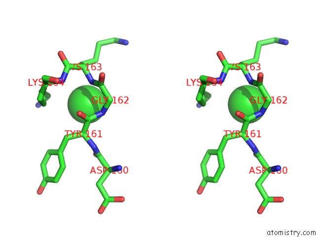
Stereo pair view
Mono view
Stereo pair view
A full contact list of Chlorine with other atoms in the Cl binding
site number 1 of The Structure of Sb-2-204-Tubulin Complex within 5.0Å range:
|
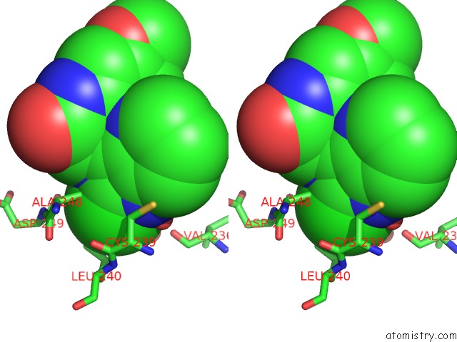
Chlorine binding site 2 out of 3 in 6n47
Go back to
Chlorine binding site 2 out
of 3 in the The Structure of Sb-2-204-Tubulin Complex
Mono view
Stereo pair view
Mono view
Stereo pair view
A full contact list of Chlorine with other atoms in the Cl binding
site number 2 of The Structure of Sb-2-204-Tubulin Complex within 5.0Å range:
|
Chlorine binding site 3 out of 3 in 6n47
Go back to
Chlorine binding site 3 out
of 3 in the The Structure of Sb-2-204-Tubulin Complex

Mono view

Stereo pair view
Mono view
Stereo pair view
A full contact list of Chlorine with other atoms in the Cl binding
site number 3 of The Structure of Sb-2-204-Tubulin Complex within 5.0Å range:
|
Reference:
K.E.Arnst,
S.Banerjee,
Y.Wang,
H.Chen,
Y.Li,
L.Yang,
W.Li,
D.D.Miller,
W.Li.
X-Ray Crystal Structure Guided Discovery and Antitumor Efficacy of Dihydroquinoxalinone As Potent Tubulin Polymerization Inhibitors. Acs Chem.Biol. 2019.
ISSN: ESSN 1554-8937
PubMed: 31714738
DOI: 10.1021/ACSCHEMBIO.9B00696
Page generated: Sun Jul 28 20:24:48 2024
ISSN: ESSN 1554-8937
PubMed: 31714738
DOI: 10.1021/ACSCHEMBIO.9B00696
Last articles
Ca in 5TACCa in 5TA0
Ca in 5TA1
Ca in 5T9Q
Ca in 5T9K
Ca in 5T9I
Ca in 5T8D
Ca in 5T9C
Ca in 5T9B
Ca in 5T91