Chlorine »
PDB 6nvh-6o0v »
6o0k »
Chlorine in PDB 6o0k: Crystal Structure of Bcl-2 with Venetoclax
Protein crystallography data
The structure of Crystal Structure of Bcl-2 with Venetoclax, PDB code: 6o0k
was solved by
R.W.Birkinshaw,
C.S.Luo,
P.M.Colman,
P.E.Czabotar,
with X-Ray Crystallography technique. A brief refinement statistics is given in the table below:
| Resolution Low / High (Å) | 43.66 / 1.62 |
| Space group | P 21 21 21 |
| Cell size a, b, c (Å), α, β, γ (°) | 33.725, 48.506, 87.316, 90.00, 90.00, 90.00 |
| R / Rfree (%) | 16.1 / 20.2 |
Chlorine Binding Sites:
The binding sites of Chlorine atom in the Crystal Structure of Bcl-2 with Venetoclax
(pdb code 6o0k). This binding sites where shown within
5.0 Angstroms radius around Chlorine atom.
In total 2 binding sites of Chlorine where determined in the Crystal Structure of Bcl-2 with Venetoclax, PDB code: 6o0k:
Jump to Chlorine binding site number: 1; 2;
In total 2 binding sites of Chlorine where determined in the Crystal Structure of Bcl-2 with Venetoclax, PDB code: 6o0k:
Jump to Chlorine binding site number: 1; 2;
Chlorine binding site 1 out of 2 in 6o0k
Go back to
Chlorine binding site 1 out
of 2 in the Crystal Structure of Bcl-2 with Venetoclax

Mono view
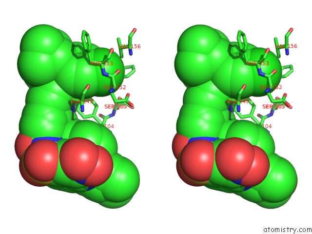
Stereo pair view
Mono view
Stereo pair view
A full contact list of Chlorine with other atoms in the Cl binding
site number 1 of Crystal Structure of Bcl-2 with Venetoclax within 5.0Å range:
|
Chlorine binding site 2 out of 2 in 6o0k
Go back to
Chlorine binding site 2 out
of 2 in the Crystal Structure of Bcl-2 with Venetoclax
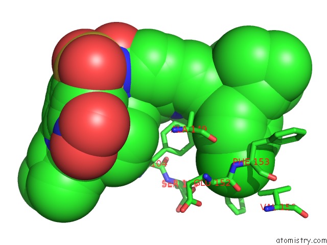
Mono view

Stereo pair view
Mono view
Stereo pair view
A full contact list of Chlorine with other atoms in the Cl binding
site number 2 of Crystal Structure of Bcl-2 with Venetoclax within 5.0Å range:
|
Reference:
R.W.Birkinshaw,
J.N.Gong,
C.S.Luo,
D.Lio,
C.A.White,
M.A.Anderson,
P.Blombery,
G.Lessene,
I.J.Majewski,
R.Thijssen,
A.W.Roberts,
D.C.S.Huang,
P.M.Colman,
P.E.Czabotar.
Structures of Bcl-2 in Complex with Venetoclax Reveal the Molecular Basis of Resistance Mutations. Nat Commun V. 10 2385 2019.
ISSN: ESSN 2041-1723
PubMed: 31160589
DOI: 10.1038/S41467-019-10363-1
Page generated: Sat Jul 12 17:36:16 2025
ISSN: ESSN 2041-1723
PubMed: 31160589
DOI: 10.1038/S41467-019-10363-1
Last articles
Fe in 2YXOFe in 2YRS
Fe in 2YXC
Fe in 2YNM
Fe in 2YVJ
Fe in 2YP1
Fe in 2YU2
Fe in 2YU1
Fe in 2YQB
Fe in 2YOO