Chlorine »
PDB 6o9g-6ok0 »
6ofs »
Chlorine in PDB 6ofs: The Crystal Structure of the Periplasmic Protease Pqql From Escherichia Coli
Protein crystallography data
The structure of The Crystal Structure of the Periplasmic Protease Pqql From Escherichia Coli, PDB code: 6ofs
was solved by
R.Grinter,
with X-Ray Crystallography technique. A brief refinement statistics is given in the table below:
| Resolution Low / High (Å) | 44.13 / 2.60 |
| Space group | P 43 21 2 |
| Cell size a, b, c (Å), α, β, γ (°) | 98.674, 98.674, 230.814, 90.00, 90.00, 90.00 |
| R / Rfree (%) | 19.9 / 24.5 |
Other elements in 6ofs:
The structure of The Crystal Structure of the Periplasmic Protease Pqql From Escherichia Coli also contains other interesting chemical elements:
| Zinc | (Zn) | 1 atom |
Chlorine Binding Sites:
The binding sites of Chlorine atom in the The Crystal Structure of the Periplasmic Protease Pqql From Escherichia Coli
(pdb code 6ofs). This binding sites where shown within
5.0 Angstroms radius around Chlorine atom.
In total 2 binding sites of Chlorine where determined in the The Crystal Structure of the Periplasmic Protease Pqql From Escherichia Coli, PDB code: 6ofs:
Jump to Chlorine binding site number: 1; 2;
In total 2 binding sites of Chlorine where determined in the The Crystal Structure of the Periplasmic Protease Pqql From Escherichia Coli, PDB code: 6ofs:
Jump to Chlorine binding site number: 1; 2;
Chlorine binding site 1 out of 2 in 6ofs
Go back to
Chlorine binding site 1 out
of 2 in the The Crystal Structure of the Periplasmic Protease Pqql From Escherichia Coli
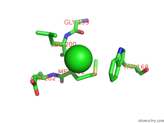
Mono view
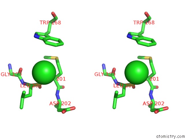
Stereo pair view
Mono view
Stereo pair view
A full contact list of Chlorine with other atoms in the Cl binding
site number 1 of The Crystal Structure of the Periplasmic Protease Pqql From Escherichia Coli within 5.0Å range:
|
Chlorine binding site 2 out of 2 in 6ofs
Go back to
Chlorine binding site 2 out
of 2 in the The Crystal Structure of the Periplasmic Protease Pqql From Escherichia Coli
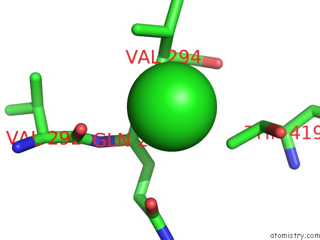
Mono view
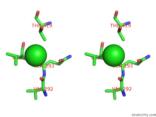
Stereo pair view
Mono view
Stereo pair view
A full contact list of Chlorine with other atoms in the Cl binding
site number 2 of The Crystal Structure of the Periplasmic Protease Pqql From Escherichia Coli within 5.0Å range:
|
Reference:
R.Grinter,
P.M.Leung,
L.C.Wijeyewickrema,
D.Littler,
S.Beckham,
R.N.Pike,
D.Walker,
C.Greening,
T.Lithgow.
Protease-Associated Import Systems Are Widespread in Gram-Negative Bacteria. Plos Genet. V. 15 08435 2019.
ISSN: ESSN 1553-7404
PubMed: 31613892
DOI: 10.1371/JOURNAL.PGEN.1008435
Page generated: Mon Jul 29 12:39:57 2024
ISSN: ESSN 1553-7404
PubMed: 31613892
DOI: 10.1371/JOURNAL.PGEN.1008435
Last articles
Zn in 9J0NZn in 9J0O
Zn in 9J0P
Zn in 9FJX
Zn in 9EKB
Zn in 9C0F
Zn in 9CAH
Zn in 9CH0
Zn in 9CH3
Zn in 9CH1