Chlorine »
PDB 6o9g-6ok0 »
6ofu »
Chlorine in PDB 6ofu: X-Ray Crystal Structure of the Ydji Aldolase From Escherichia Coli K12
Protein crystallography data
The structure of X-Ray Crystal Structure of the Ydji Aldolase From Escherichia Coli K12, PDB code: 6ofu
was solved by
B.J.Dopkins,
J.B.Thoden,
J.P.Huddleston,
T.Narindoshvili,
B.Fose,
F.M.Rachel,
H.M.Holden,
with X-Ray Crystallography technique. A brief refinement statistics is given in the table below:
| Resolution Low / High (Å) | 34.74 / 1.75 |
| Space group | P 1 21 1 |
| Cell size a, b, c (Å), α, β, γ (°) | 80.763, 85.663, 84.268, 90.00, 109.88, 90.00 |
| R / Rfree (%) | 14.1 / 18.2 |
Other elements in 6ofu:
The structure of X-Ray Crystal Structure of the Ydji Aldolase From Escherichia Coli K12 also contains other interesting chemical elements:
| Zinc | (Zn) | 5 atoms |
Chlorine Binding Sites:
The binding sites of Chlorine atom in the X-Ray Crystal Structure of the Ydji Aldolase From Escherichia Coli K12
(pdb code 6ofu). This binding sites where shown within
5.0 Angstroms radius around Chlorine atom.
In total 2 binding sites of Chlorine where determined in the X-Ray Crystal Structure of the Ydji Aldolase From Escherichia Coli K12, PDB code: 6ofu:
Jump to Chlorine binding site number: 1; 2;
In total 2 binding sites of Chlorine where determined in the X-Ray Crystal Structure of the Ydji Aldolase From Escherichia Coli K12, PDB code: 6ofu:
Jump to Chlorine binding site number: 1; 2;
Chlorine binding site 1 out of 2 in 6ofu
Go back to
Chlorine binding site 1 out
of 2 in the X-Ray Crystal Structure of the Ydji Aldolase From Escherichia Coli K12
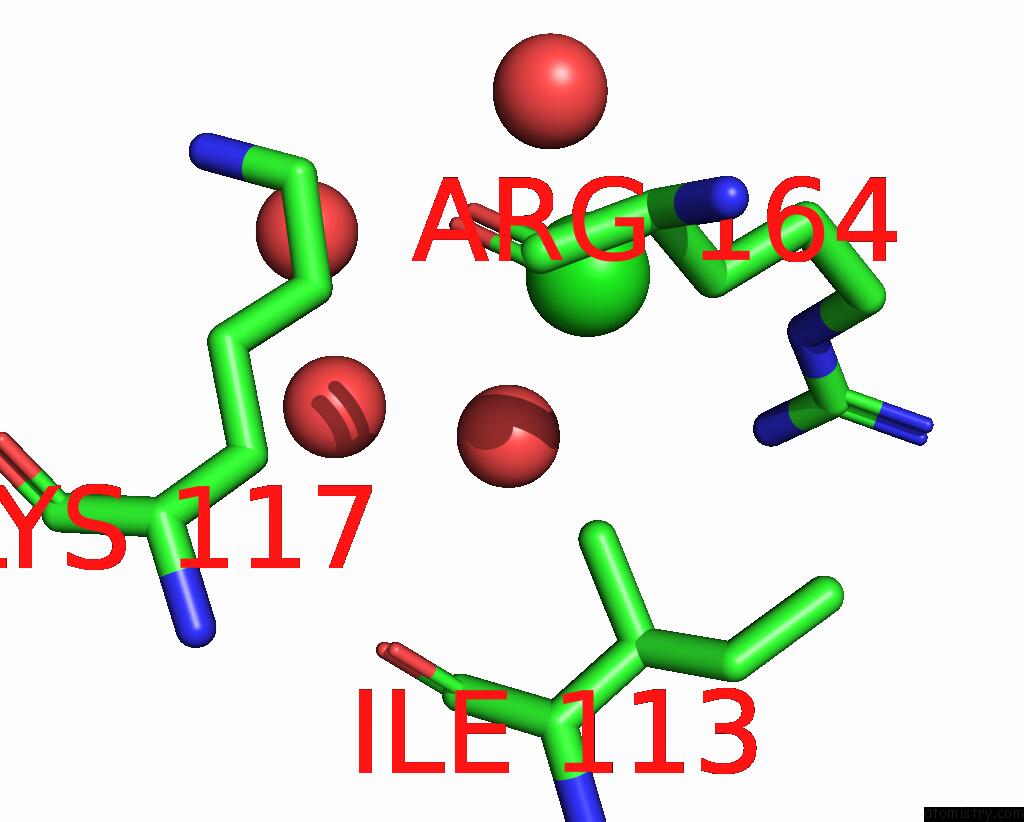
Mono view
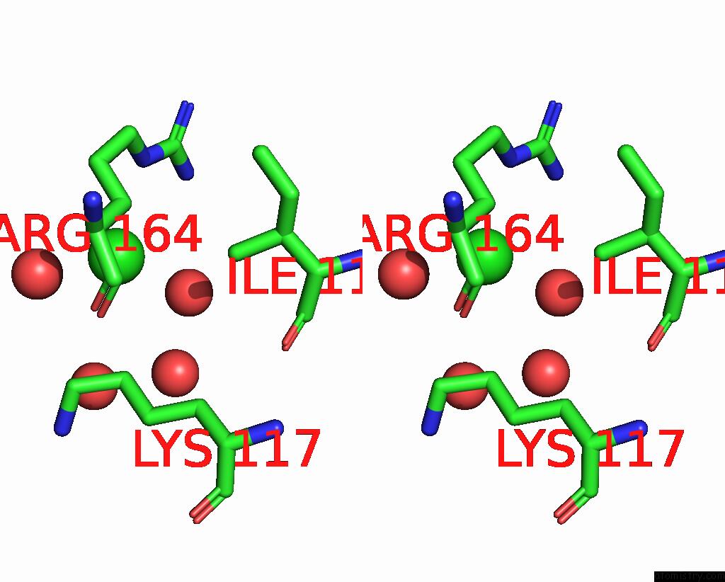
Stereo pair view
Mono view
Stereo pair view
A full contact list of Chlorine with other atoms in the Cl binding
site number 1 of X-Ray Crystal Structure of the Ydji Aldolase From Escherichia Coli K12 within 5.0Å range:
|
Chlorine binding site 2 out of 2 in 6ofu
Go back to
Chlorine binding site 2 out
of 2 in the X-Ray Crystal Structure of the Ydji Aldolase From Escherichia Coli K12
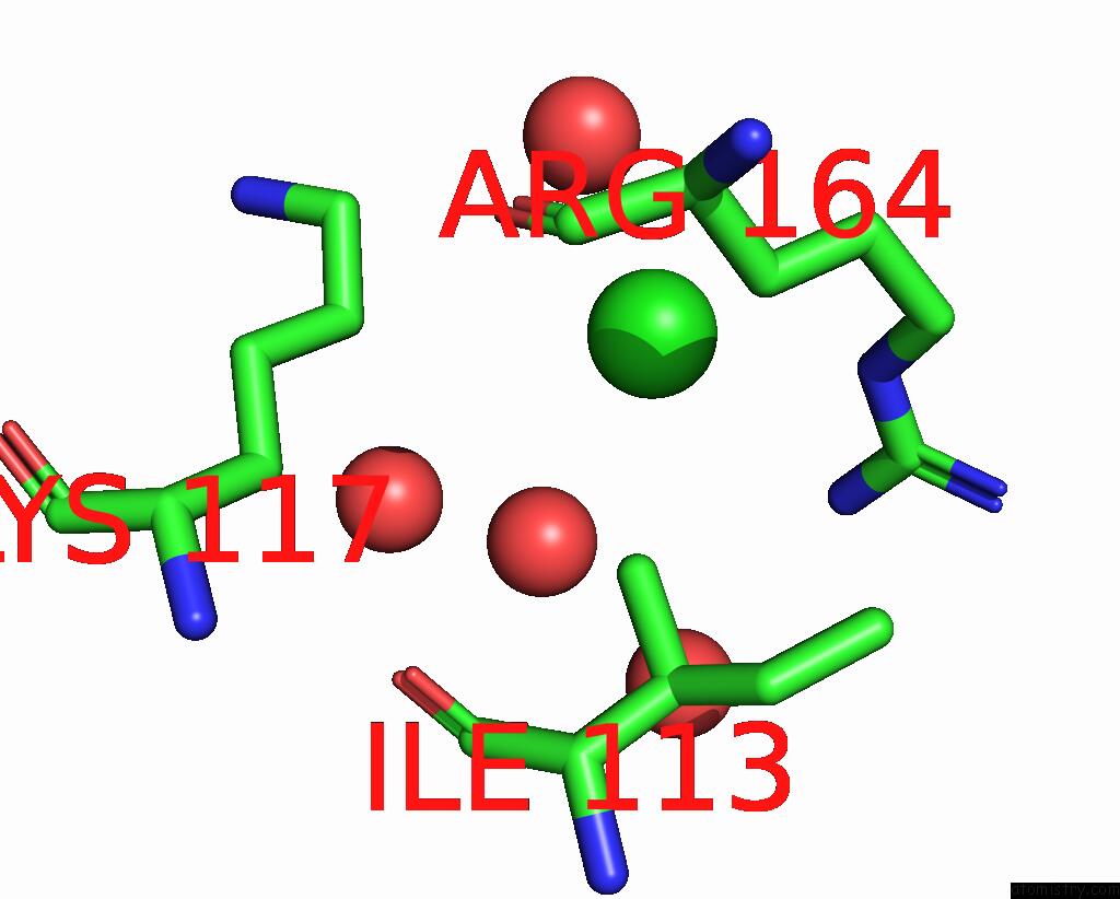
Mono view
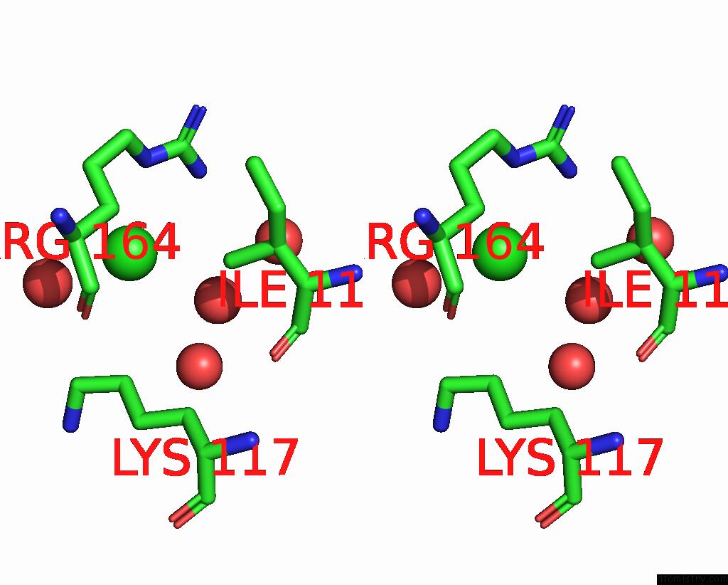
Stereo pair view
Mono view
Stereo pair view
A full contact list of Chlorine with other atoms in the Cl binding
site number 2 of X-Ray Crystal Structure of the Ydji Aldolase From Escherichia Coli K12 within 5.0Å range:
|
Reference:
J.P.Huddleston,
J.B.Thoden,
B.J.Dopkins,
T.Narindoshvili,
B.J.Fose,
H.M.Holden,
F.M.Raushel.
Structural and Functional Characterization of Ydji, An Aldolase of Unknown Specificity Inescherichia COLIK12. Biochemistry V. 58 3340 2019.
ISSN: ISSN 0006-2960
PubMed: 31322866
DOI: 10.1021/ACS.BIOCHEM.9B00326
Page generated: Mon Jul 29 12:40:11 2024
ISSN: ISSN 0006-2960
PubMed: 31322866
DOI: 10.1021/ACS.BIOCHEM.9B00326
Last articles
Zn in 9J0NZn in 9J0O
Zn in 9J0P
Zn in 9FJX
Zn in 9EKB
Zn in 9C0F
Zn in 9CAH
Zn in 9CH0
Zn in 9CH3
Zn in 9CH1