Chlorine »
PDB 6ok1-6osu »
6om5 »
Chlorine in PDB 6om5: Structure of A Haemophore From Haemophilus Haemolyticus
Protein crystallography data
The structure of Structure of A Haemophore From Haemophilus Haemolyticus, PDB code: 6om5
was solved by
M.Torrado,
J.L.Walshe,
J.P.Mackay,
J.M.Guss,
D.A.Gell,
with X-Ray Crystallography technique. A brief refinement statistics is given in the table below:
| Resolution Low / High (Å) | 41.49 / 1.60 |
| Space group | P 32 2 1 |
| Cell size a, b, c (Å), α, β, γ (°) | 91.043, 91.043, 97.484, 90.00, 90.00, 120.00 |
| R / Rfree (%) | 17.1 / 18.4 |
Other elements in 6om5:
The structure of Structure of A Haemophore From Haemophilus Haemolyticus also contains other interesting chemical elements:
| Iron | (Fe) | 1 atom |
Chlorine Binding Sites:
The binding sites of Chlorine atom in the Structure of A Haemophore From Haemophilus Haemolyticus
(pdb code 6om5). This binding sites where shown within
5.0 Angstroms radius around Chlorine atom.
In total 2 binding sites of Chlorine where determined in the Structure of A Haemophore From Haemophilus Haemolyticus, PDB code: 6om5:
Jump to Chlorine binding site number: 1; 2;
In total 2 binding sites of Chlorine where determined in the Structure of A Haemophore From Haemophilus Haemolyticus, PDB code: 6om5:
Jump to Chlorine binding site number: 1; 2;
Chlorine binding site 1 out of 2 in 6om5
Go back to
Chlorine binding site 1 out
of 2 in the Structure of A Haemophore From Haemophilus Haemolyticus
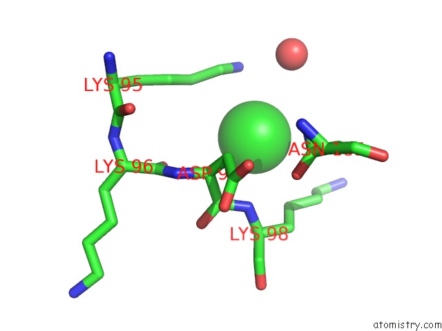
Mono view
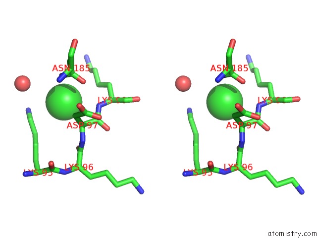
Stereo pair view
Mono view
Stereo pair view
A full contact list of Chlorine with other atoms in the Cl binding
site number 1 of Structure of A Haemophore From Haemophilus Haemolyticus within 5.0Å range:
|
Chlorine binding site 2 out of 2 in 6om5
Go back to
Chlorine binding site 2 out
of 2 in the Structure of A Haemophore From Haemophilus Haemolyticus
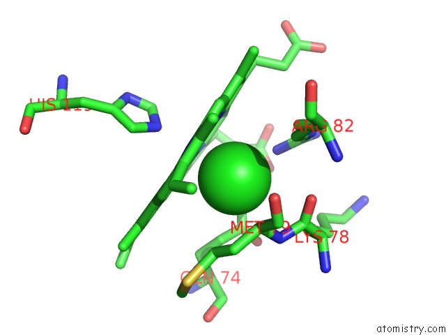
Mono view
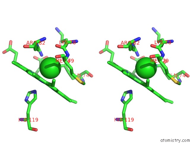
Stereo pair view
Mono view
Stereo pair view
A full contact list of Chlorine with other atoms in the Cl binding
site number 2 of Structure of A Haemophore From Haemophilus Haemolyticus within 5.0Å range:
|
Reference:
R.D.Latham,
M.Torrado,
B.Atto,
J.L.Walshe,
R.Wilson,
J.M.Guss,
J.P.Mackay,
S.Tristram,
D.A.Gell.
A Heme-Binding Protein Produced By Haemophilus Haemolyticus Inhibits Non-Typeable Haemophilus Influenzae. Mol.Microbiol. 2019.
ISSN: ESSN 1365-2958
PubMed: 31742788
DOI: 10.1111/MMI.14426
Page generated: Mon Jul 29 12:43:49 2024
ISSN: ESSN 1365-2958
PubMed: 31742788
DOI: 10.1111/MMI.14426
Last articles
Zn in 9J0NZn in 9J0O
Zn in 9J0P
Zn in 9FJX
Zn in 9EKB
Zn in 9C0F
Zn in 9CAH
Zn in 9CH0
Zn in 9CH3
Zn in 9CH1