Chlorine »
PDB 6otc-6p2k »
6ov8 »
Chlorine in PDB 6ov8: 2.6 Angstrom Resolution Crystal Structure of Aminopeptidase B From Escherichia Coli Str. K-12 Substr. MG1655
Enzymatic activity of 2.6 Angstrom Resolution Crystal Structure of Aminopeptidase B From Escherichia Coli Str. K-12 Substr. MG1655
All present enzymatic activity of 2.6 Angstrom Resolution Crystal Structure of Aminopeptidase B From Escherichia Coli Str. K-12 Substr. MG1655:
3.4.11.23;
3.4.11.23;
Protein crystallography data
The structure of 2.6 Angstrom Resolution Crystal Structure of Aminopeptidase B From Escherichia Coli Str. K-12 Substr. MG1655, PDB code: 6ov8
was solved by
G.Minasov,
L.Shuvalova,
Z.Wawrzak,
O.Kiryukhina,
S.Grimshaw,
K.Kwon,
K.J.F.Satchell,
Center For Structural Genomics Of Infectious Diseases(Csgid),
with X-Ray Crystallography technique. A brief refinement statistics is given in the table below:
| Resolution Low / High (Å) | 29.76 / 2.61 |
| Space group | P 21 21 21 |
| Cell size a, b, c (Å), α, β, γ (°) | 114.857, 148.190, 165.010, 90.00, 90.00, 90.00 |
| R / Rfree (%) | 18.1 / 23.8 |
Other elements in 6ov8:
The structure of 2.6 Angstrom Resolution Crystal Structure of Aminopeptidase B From Escherichia Coli Str. K-12 Substr. MG1655 also contains other interesting chemical elements:
| Manganese | (Mn) | 6 atoms |
| Zinc | (Zn) | 6 atoms |
Chlorine Binding Sites:
Pages:
>>> Page 1 <<< Page 2, Binding sites: 11 - 20; Page 3, Binding sites: 21 - 21;Binding sites:
The binding sites of Chlorine atom in the 2.6 Angstrom Resolution Crystal Structure of Aminopeptidase B From Escherichia Coli Str. K-12 Substr. MG1655 (pdb code 6ov8). This binding sites where shown within 5.0 Angstroms radius around Chlorine atom.In total 21 binding sites of Chlorine where determined in the 2.6 Angstrom Resolution Crystal Structure of Aminopeptidase B From Escherichia Coli Str. K-12 Substr. MG1655, PDB code: 6ov8:
Jump to Chlorine binding site number: 1; 2; 3; 4; 5; 6; 7; 8; 9; 10;
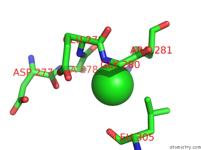
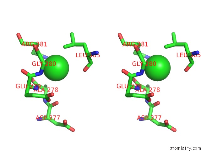
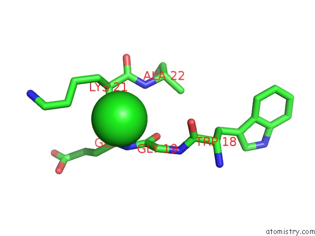
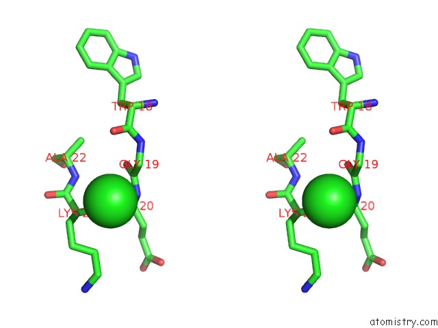
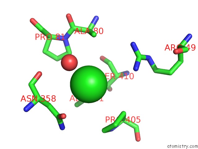
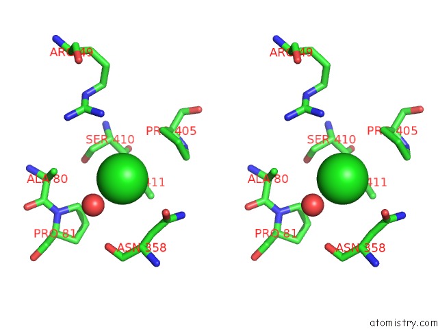
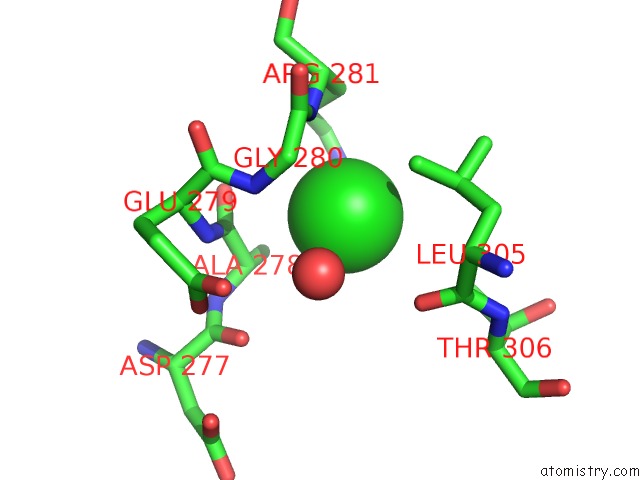
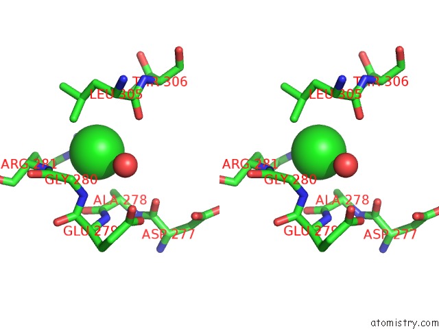
Chlorine binding site 1 out of 21 in 6ov8
Go back to
Chlorine binding site 1 out
of 21 in the 2.6 Angstrom Resolution Crystal Structure of Aminopeptidase B From Escherichia Coli Str. K-12 Substr. MG1655
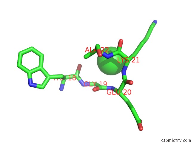
Mono view
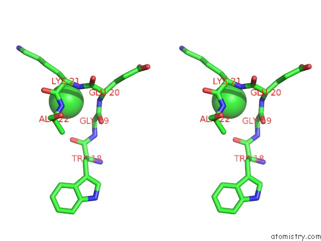
Stereo pair view
Mono view
Stereo pair view
A full contact list of Chlorine with other atoms in the Cl binding
site number 1 of 2.6 Angstrom Resolution Crystal Structure of Aminopeptidase B From Escherichia Coli Str. K-12 Substr. MG1655 within 5.0Å range:
|
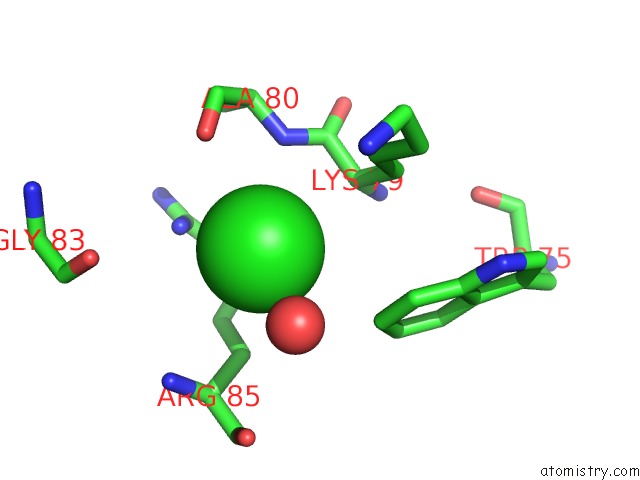
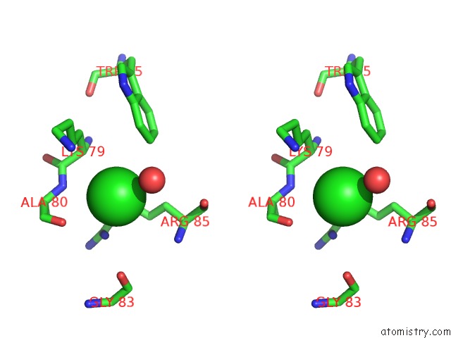
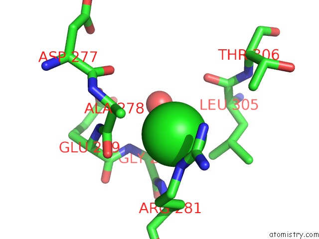
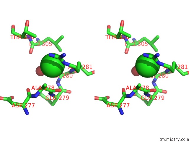
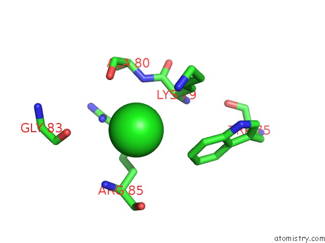
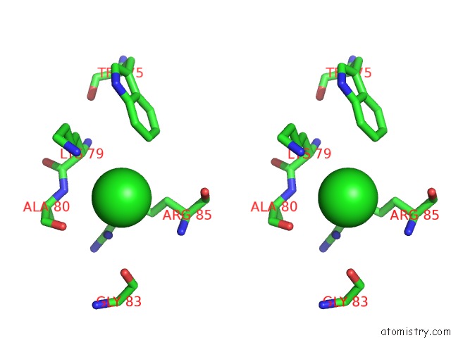
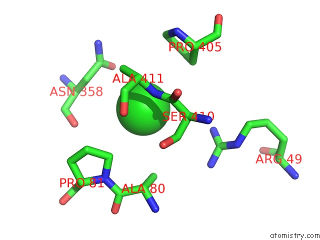
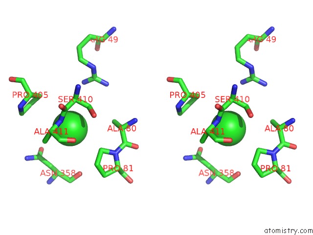
Chlorine binding site 2 out of 21 in 6ov8
Go back to
Chlorine binding site 2 out
of 21 in the 2.6 Angstrom Resolution Crystal Structure of Aminopeptidase B From Escherichia Coli Str. K-12 Substr. MG1655
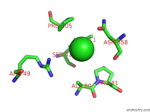
Mono view
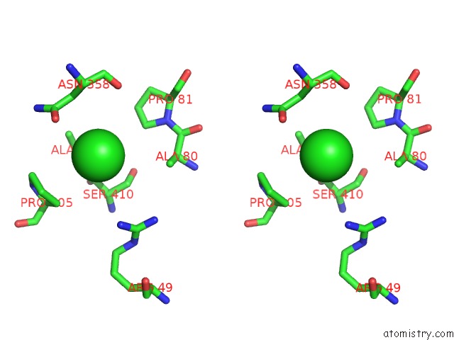
Stereo pair view
Mono view
Stereo pair view
A full contact list of Chlorine with other atoms in the Cl binding
site number 2 of 2.6 Angstrom Resolution Crystal Structure of Aminopeptidase B From Escherichia Coli Str. K-12 Substr. MG1655 within 5.0Å range:
|
Chlorine binding site 3 out of 21 in 6ov8
Go back to
Chlorine binding site 3 out
of 21 in the 2.6 Angstrom Resolution Crystal Structure of Aminopeptidase B From Escherichia Coli Str. K-12 Substr. MG1655
Mono view
Stereo pair view
Mono view
Stereo pair view
A full contact list of Chlorine with other atoms in the Cl binding
site number 3 of 2.6 Angstrom Resolution Crystal Structure of Aminopeptidase B From Escherichia Coli Str. K-12 Substr. MG1655 within 5.0Å range:
|
Chlorine binding site 4 out of 21 in 6ov8
Go back to
Chlorine binding site 4 out
of 21 in the 2.6 Angstrom Resolution Crystal Structure of Aminopeptidase B From Escherichia Coli Str. K-12 Substr. MG1655
Mono view
Stereo pair view
Mono view
Stereo pair view
A full contact list of Chlorine with other atoms in the Cl binding
site number 4 of 2.6 Angstrom Resolution Crystal Structure of Aminopeptidase B From Escherichia Coli Str. K-12 Substr. MG1655 within 5.0Å range:
|
Chlorine binding site 5 out of 21 in 6ov8
Go back to
Chlorine binding site 5 out
of 21 in the 2.6 Angstrom Resolution Crystal Structure of Aminopeptidase B From Escherichia Coli Str. K-12 Substr. MG1655
Mono view
Stereo pair view
Mono view
Stereo pair view
A full contact list of Chlorine with other atoms in the Cl binding
site number 5 of 2.6 Angstrom Resolution Crystal Structure of Aminopeptidase B From Escherichia Coli Str. K-12 Substr. MG1655 within 5.0Å range:
|
Chlorine binding site 6 out of 21 in 6ov8
Go back to
Chlorine binding site 6 out
of 21 in the 2.6 Angstrom Resolution Crystal Structure of Aminopeptidase B From Escherichia Coli Str. K-12 Substr. MG1655
Mono view
Stereo pair view
Mono view
Stereo pair view
A full contact list of Chlorine with other atoms in the Cl binding
site number 6 of 2.6 Angstrom Resolution Crystal Structure of Aminopeptidase B From Escherichia Coli Str. K-12 Substr. MG1655 within 5.0Å range:
|
Chlorine binding site 7 out of 21 in 6ov8
Go back to
Chlorine binding site 7 out
of 21 in the 2.6 Angstrom Resolution Crystal Structure of Aminopeptidase B From Escherichia Coli Str. K-12 Substr. MG1655
Mono view
Stereo pair view
Mono view
Stereo pair view
A full contact list of Chlorine with other atoms in the Cl binding
site number 7 of 2.6 Angstrom Resolution Crystal Structure of Aminopeptidase B From Escherichia Coli Str. K-12 Substr. MG1655 within 5.0Å range:
|
Chlorine binding site 8 out of 21 in 6ov8
Go back to
Chlorine binding site 8 out
of 21 in the 2.6 Angstrom Resolution Crystal Structure of Aminopeptidase B From Escherichia Coli Str. K-12 Substr. MG1655
Mono view
Stereo pair view
Mono view
Stereo pair view
A full contact list of Chlorine with other atoms in the Cl binding
site number 8 of 2.6 Angstrom Resolution Crystal Structure of Aminopeptidase B From Escherichia Coli Str. K-12 Substr. MG1655 within 5.0Å range:
|
Chlorine binding site 9 out of 21 in 6ov8
Go back to
Chlorine binding site 9 out
of 21 in the 2.6 Angstrom Resolution Crystal Structure of Aminopeptidase B From Escherichia Coli Str. K-12 Substr. MG1655
Mono view
Stereo pair view
Mono view
Stereo pair view
A full contact list of Chlorine with other atoms in the Cl binding
site number 9 of 2.6 Angstrom Resolution Crystal Structure of Aminopeptidase B From Escherichia Coli Str. K-12 Substr. MG1655 within 5.0Å range:
|
Chlorine binding site 10 out of 21 in 6ov8
Go back to
Chlorine binding site 10 out
of 21 in the 2.6 Angstrom Resolution Crystal Structure of Aminopeptidase B From Escherichia Coli Str. K-12 Substr. MG1655
Mono view
Stereo pair view
Mono view
Stereo pair view
A full contact list of Chlorine with other atoms in the Cl binding
site number 10 of 2.6 Angstrom Resolution Crystal Structure of Aminopeptidase B From Escherichia Coli Str. K-12 Substr. MG1655 within 5.0Å range:
|
Reference:
G.Minasov,
L.Shuvalova,
Z.Wawrzak,
O.Kiryukhina,
S.Grimshaw,
K.Kwon,
K.J.F.Satchell,
Center For Structural Genomics Of Infectious Diseases(Csgid).
2.6 Angstrom Resolution Crystal Structure of Aminopeptidase B From Escherichia Coli Str. K-12 Substr. MG1655. To Be Published.
Page generated: Mon Jul 29 12:53:51 2024
Last articles
Ca in 5SB6Ca in 5SB5
Ca in 5SB3
Ca in 5SB4
Ca in 5S9M
Ca in 5S9N
Ca in 5S9L
Ca in 5S8Q
Ca in 5S8P
Ca in 5S8O