Chlorine »
PDB 6p2l-6p8z »
6p2n »
Chlorine in PDB 6p2n: Crystal Structure of Paenibacillus Graminis GH74 (PGGH74)
Protein crystallography data
The structure of Crystal Structure of Paenibacillus Graminis GH74 (PGGH74), PDB code: 6p2n
was solved by
P.J.Stogios,
with X-Ray Crystallography technique. A brief refinement statistics is given in the table below:
| Resolution Low / High (Å) | 30.45 / 1.35 |
| Space group | P 21 21 21 |
| Cell size a, b, c (Å), α, β, γ (°) | 57.494, 86.214, 130.197, 90.00, 90.00, 90.00 |
| R / Rfree (%) | 14.9 / 18 |
Other elements in 6p2n:
The structure of Crystal Structure of Paenibacillus Graminis GH74 (PGGH74) also contains other interesting chemical elements:
| Magnesium | (Mg) | 1 atom |
Chlorine Binding Sites:
The binding sites of Chlorine atom in the Crystal Structure of Paenibacillus Graminis GH74 (PGGH74)
(pdb code 6p2n). This binding sites where shown within
5.0 Angstroms radius around Chlorine atom.
In total 2 binding sites of Chlorine where determined in the Crystal Structure of Paenibacillus Graminis GH74 (PGGH74), PDB code: 6p2n:
Jump to Chlorine binding site number: 1; 2;
In total 2 binding sites of Chlorine where determined in the Crystal Structure of Paenibacillus Graminis GH74 (PGGH74), PDB code: 6p2n:
Jump to Chlorine binding site number: 1; 2;
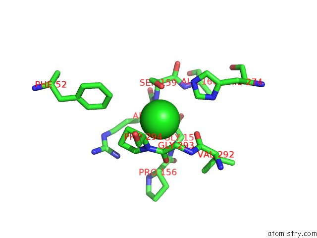
Chlorine binding site 1 out of 2 in 6p2n
Go back to
Chlorine binding site 1 out
of 2 in the Crystal Structure of Paenibacillus Graminis GH74 (PGGH74)
Mono view
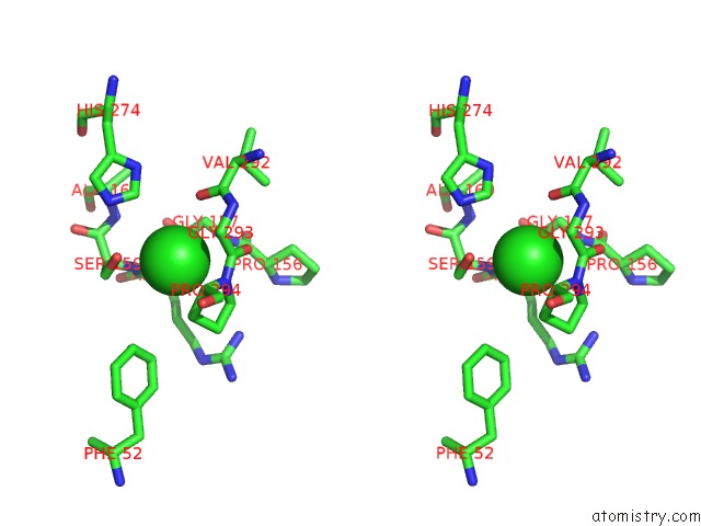
Stereo pair view
Mono view
Stereo pair view
A full contact list of Chlorine with other atoms in the Cl binding
site number 1 of Crystal Structure of Paenibacillus Graminis GH74 (PGGH74) within 5.0Å range:
|
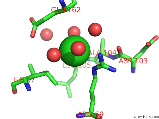
Chlorine binding site 2 out of 2 in 6p2n
Go back to
Chlorine binding site 2 out
of 2 in the Crystal Structure of Paenibacillus Graminis GH74 (PGGH74)
Mono view
Stereo pair view
Mono view
Stereo pair view
A full contact list of Chlorine with other atoms in the Cl binding
site number 2 of Crystal Structure of Paenibacillus Graminis GH74 (PGGH74) within 5.0Å range:
|
Reference:
G.Arnal,
P.J.Stogios,
J.Asohan,
M.A.Attia,
T.Skarina,
A.H.Viborg,
B.Henrissat,
A.Savchenko,
H.Brumer.
Substrate Specificity, Regiospecificity, and Processivity in Glycoside Hydrolase Family 74. J.Biol.Chem. V. 294 13233 2019.
ISSN: ESSN 1083-351X
PubMed: 31324716
DOI: 10.1074/JBC.RA119.009861
Page generated: Sat Jul 12 18:09:47 2025
ISSN: ESSN 1083-351X
PubMed: 31324716
DOI: 10.1074/JBC.RA119.009861
Last articles
F in 4HT0F in 4HNA
F in 4HPX
F in 4HQH
F in 4HNS
F in 4HPJ
F in 4HN4
F in 4HJX
F in 4HLH
F in 4HL4