Chlorine »
PDB 6p90-6pil »
6pbb »
Chlorine in PDB 6pbb: Crystal Structure of Hen Egg White Lysozyme in Complex with I3C
Enzymatic activity of Crystal Structure of Hen Egg White Lysozyme in Complex with I3C
All present enzymatic activity of Crystal Structure of Hen Egg White Lysozyme in Complex with I3C:
3.2.1.17;
3.2.1.17;
Protein crystallography data
The structure of Crystal Structure of Hen Egg White Lysozyme in Complex with I3C, PDB code: 6pbb
was solved by
J.Q.Truong,
with X-Ray Crystallography technique. A brief refinement statistics is given in the table below:
| Resolution Low / High (Å) | 34.51 / 1.89 |
| Space group | P 43 21 2 |
| Cell size a, b, c (Å), α, β, γ (°) | 77.160, 77.160, 38.201, 90.00, 90.00, 90.00 |
| R / Rfree (%) | 19.7 / 24 |
Other elements in 6pbb:
The structure of Crystal Structure of Hen Egg White Lysozyme in Complex with I3C also contains other interesting chemical elements:
| Iodine | (I) | 12 atoms |
Chlorine Binding Sites:
The binding sites of Chlorine atom in the Crystal Structure of Hen Egg White Lysozyme in Complex with I3C
(pdb code 6pbb). This binding sites where shown within
5.0 Angstroms radius around Chlorine atom.
In total only one binding site of Chlorine was determined in the Crystal Structure of Hen Egg White Lysozyme in Complex with I3C, PDB code: 6pbb:
In total only one binding site of Chlorine was determined in the Crystal Structure of Hen Egg White Lysozyme in Complex with I3C, PDB code: 6pbb:
Chlorine binding site 1 out of 1 in 6pbb
Go back to
Chlorine binding site 1 out
of 1 in the Crystal Structure of Hen Egg White Lysozyme in Complex with I3C
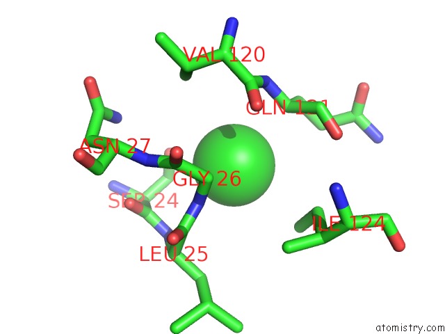
Mono view
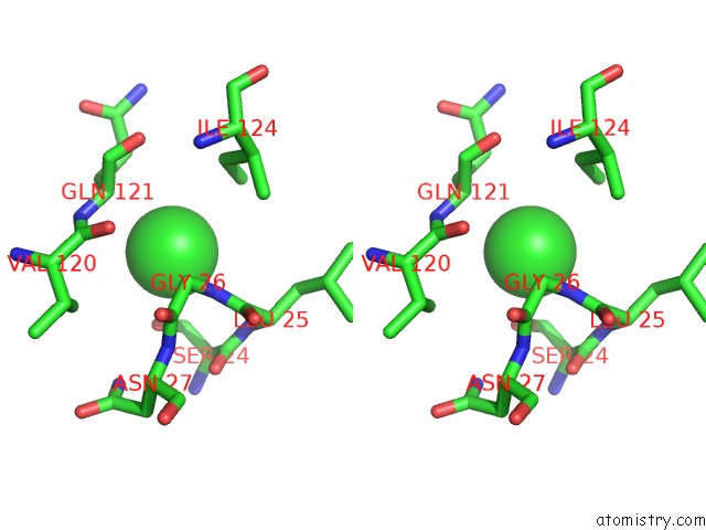
Stereo pair view
Mono view
Stereo pair view
A full contact list of Chlorine with other atoms in the Cl binding
site number 1 of Crystal Structure of Hen Egg White Lysozyme in Complex with I3C within 5.0Å range:
|
Reference:
J.Q.Truong,
S.Panjikar,
L.Shearwin-Whyatt,
J.B.Bruning,
K.E.Shearwin.
Combining Random Microseed Matrix Screening and the Magic Triangle For the Efficient Structure Solution of A Potential Lysin From Bacteriophage P68. Acta Crystallogr D Struct V. 75 670 2019BIOL.
ISSN: ISSN 2059-7983
PubMed: 31282476
DOI: 10.1107/S2059798319009008
Page generated: Mon Jul 29 13:16:49 2024
ISSN: ISSN 2059-7983
PubMed: 31282476
DOI: 10.1107/S2059798319009008
Last articles
Zn in 9J0NZn in 9J0O
Zn in 9J0P
Zn in 9FJX
Zn in 9EKB
Zn in 9C0F
Zn in 9CAH
Zn in 9CH0
Zn in 9CH3
Zn in 9CH1