Chlorine »
PDB 6p90-6pil »
6pcb »
Chlorine in PDB 6pcb: Crystal Structure of Beta-Ketoadipyl-Coa Thiolase Mutant (H356A) in Complex with Coa
Enzymatic activity of Crystal Structure of Beta-Ketoadipyl-Coa Thiolase Mutant (H356A) in Complex with Coa
All present enzymatic activity of Crystal Structure of Beta-Ketoadipyl-Coa Thiolase Mutant (H356A) in Complex with Coa:
2.3.1.16; 2.3.1.174;
2.3.1.16; 2.3.1.174;
Protein crystallography data
The structure of Crystal Structure of Beta-Ketoadipyl-Coa Thiolase Mutant (H356A) in Complex with Coa, PDB code: 6pcb
was solved by
B.Sukritee,
S.Panjikar,
with X-Ray Crystallography technique. A brief refinement statistics is given in the table below:
| Resolution Low / High (Å) | 20.00 / 1.61 |
| Space group | P 21 21 21 |
| Cell size a, b, c (Å), α, β, γ (°) | 110.487, 115.958, 128.866, 90.00, 90.00, 90.00 |
| R / Rfree (%) | 16.1 / 19.6 |
Chlorine Binding Sites:
The binding sites of Chlorine atom in the Crystal Structure of Beta-Ketoadipyl-Coa Thiolase Mutant (H356A) in Complex with Coa
(pdb code 6pcb). This binding sites where shown within
5.0 Angstroms radius around Chlorine atom.
In total 5 binding sites of Chlorine where determined in the Crystal Structure of Beta-Ketoadipyl-Coa Thiolase Mutant (H356A) in Complex with Coa, PDB code: 6pcb:
Jump to Chlorine binding site number: 1; 2; 3; 4; 5;
In total 5 binding sites of Chlorine where determined in the Crystal Structure of Beta-Ketoadipyl-Coa Thiolase Mutant (H356A) in Complex with Coa, PDB code: 6pcb:
Jump to Chlorine binding site number: 1; 2; 3; 4; 5;
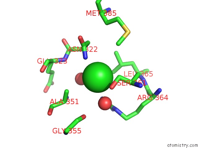
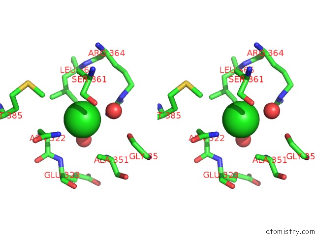
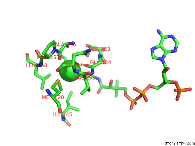
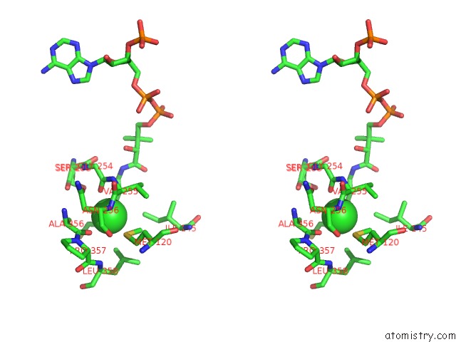
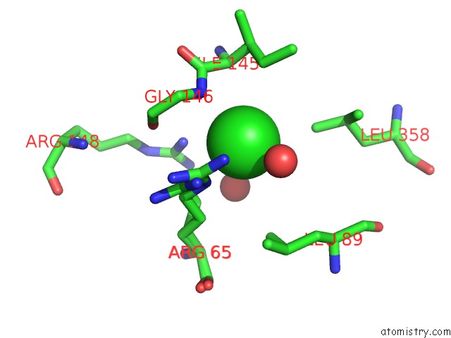
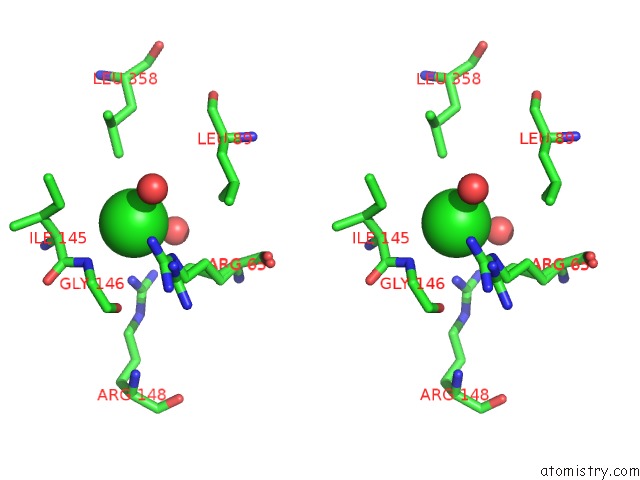
Chlorine binding site 1 out of 5 in 6pcb
Go back to
Chlorine binding site 1 out
of 5 in the Crystal Structure of Beta-Ketoadipyl-Coa Thiolase Mutant (H356A) in Complex with Coa
Mono view
Stereo pair view
Mono view
Stereo pair view
A full contact list of Chlorine with other atoms in the Cl binding
site number 1 of Crystal Structure of Beta-Ketoadipyl-Coa Thiolase Mutant (H356A) in Complex with Coa within 5.0Å range:
|
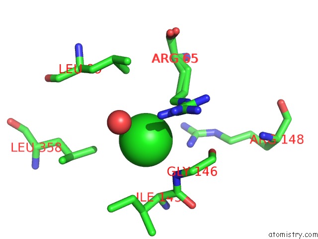
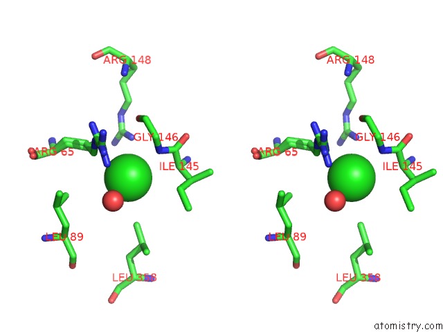
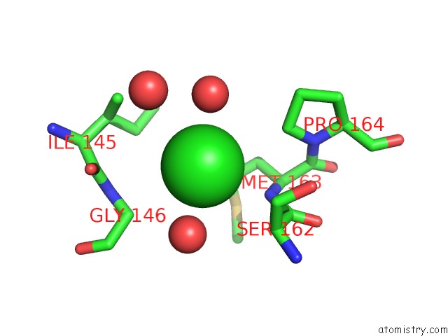
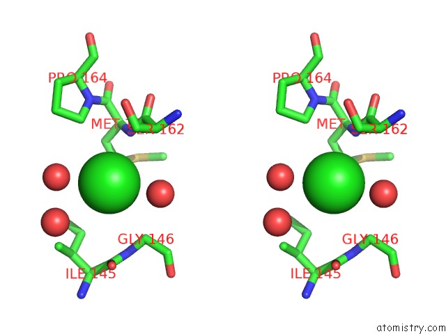
Chlorine binding site 2 out of 5 in 6pcb
Go back to
Chlorine binding site 2 out
of 5 in the Crystal Structure of Beta-Ketoadipyl-Coa Thiolase Mutant (H356A) in Complex with Coa
Mono view
Stereo pair view
Mono view
Stereo pair view
A full contact list of Chlorine with other atoms in the Cl binding
site number 2 of Crystal Structure of Beta-Ketoadipyl-Coa Thiolase Mutant (H356A) in Complex with Coa within 5.0Å range:
|
Chlorine binding site 3 out of 5 in 6pcb
Go back to
Chlorine binding site 3 out
of 5 in the Crystal Structure of Beta-Ketoadipyl-Coa Thiolase Mutant (H356A) in Complex with Coa
Mono view
Stereo pair view
Mono view
Stereo pair view
A full contact list of Chlorine with other atoms in the Cl binding
site number 3 of Crystal Structure of Beta-Ketoadipyl-Coa Thiolase Mutant (H356A) in Complex with Coa within 5.0Å range:
|
Chlorine binding site 4 out of 5 in 6pcb
Go back to
Chlorine binding site 4 out
of 5 in the Crystal Structure of Beta-Ketoadipyl-Coa Thiolase Mutant (H356A) in Complex with Coa
Mono view
Stereo pair view
Mono view
Stereo pair view
A full contact list of Chlorine with other atoms in the Cl binding
site number 4 of Crystal Structure of Beta-Ketoadipyl-Coa Thiolase Mutant (H356A) in Complex with Coa within 5.0Å range:
|
Chlorine binding site 5 out of 5 in 6pcb
Go back to
Chlorine binding site 5 out
of 5 in the Crystal Structure of Beta-Ketoadipyl-Coa Thiolase Mutant (H356A) in Complex with Coa
Mono view
Stereo pair view
Mono view
Stereo pair view
A full contact list of Chlorine with other atoms in the Cl binding
site number 5 of Crystal Structure of Beta-Ketoadipyl-Coa Thiolase Mutant (H356A) in Complex with Coa within 5.0Å range:
|
Reference:
S.Bhaskar,
D.Steer,
R.Anand,
S.Panjikar.
Structural Basis For Differentiation Between Two Classes of Thiolase: Degradative Vs Biosynthetic Thiolase J Struct Biol X 2020.
ISSN: ESSN 2590-1524
DOI: 10.1016/J.YJSBX.2019.100018
Page generated: Mon Jul 29 13:18:06 2024
ISSN: ESSN 2590-1524
DOI: 10.1016/J.YJSBX.2019.100018
Last articles
Zn in 9J0NZn in 9J0O
Zn in 9J0P
Zn in 9FJX
Zn in 9EKB
Zn in 9C0F
Zn in 9CAH
Zn in 9CH0
Zn in 9CH3
Zn in 9CH1