Chlorine »
PDB 6p90-6pil »
6pft »
Chlorine in PDB 6pft: RSEGFP2 with A Chlorinated Chromophore in the Non-Fluorescent Off- State
Protein crystallography data
The structure of RSEGFP2 with A Chlorinated Chromophore in the Non-Fluorescent Off- State, PDB code: 6pft
was solved by
J.Chang,
M.G.Romei,
S.G.Boxer,
with X-Ray Crystallography technique. A brief refinement statistics is given in the table below:
| Resolution Low / High (Å) | 34.31 / 1.45 |
| Space group | P 21 21 21 |
| Cell size a, b, c (Å), α, β, γ (°) | 51.014, 62.716, 68.619, 90.00, 90.00, 90.00 |
| R / Rfree (%) | 15 / 17.6 |
Chlorine Binding Sites:
The binding sites of Chlorine atom in the RSEGFP2 with A Chlorinated Chromophore in the Non-Fluorescent Off- State
(pdb code 6pft). This binding sites where shown within
5.0 Angstroms radius around Chlorine atom.
In total only one binding site of Chlorine was determined in the RSEGFP2 with A Chlorinated Chromophore in the Non-Fluorescent Off- State, PDB code: 6pft:
In total only one binding site of Chlorine was determined in the RSEGFP2 with A Chlorinated Chromophore in the Non-Fluorescent Off- State, PDB code: 6pft:
Chlorine binding site 1 out of 1 in 6pft
Go back to
Chlorine binding site 1 out
of 1 in the RSEGFP2 with A Chlorinated Chromophore in the Non-Fluorescent Off- State
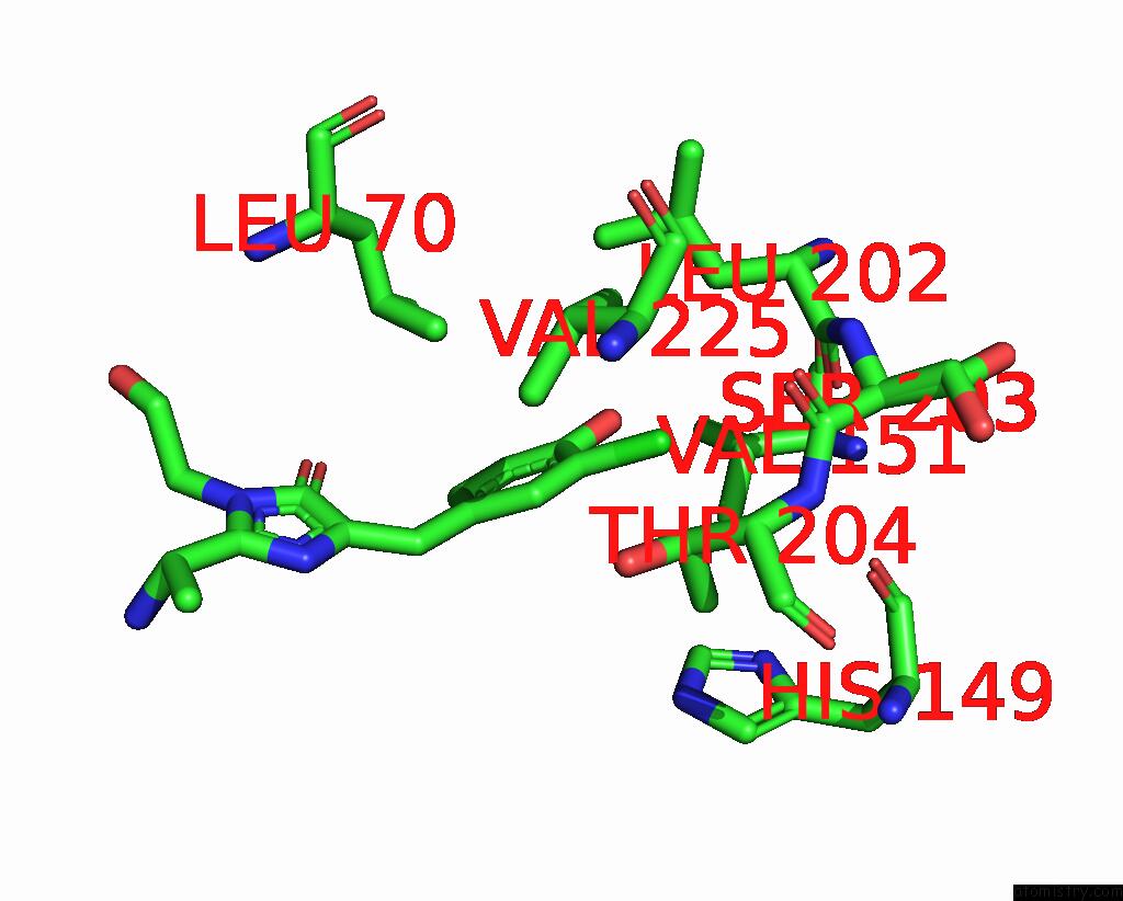
Mono view
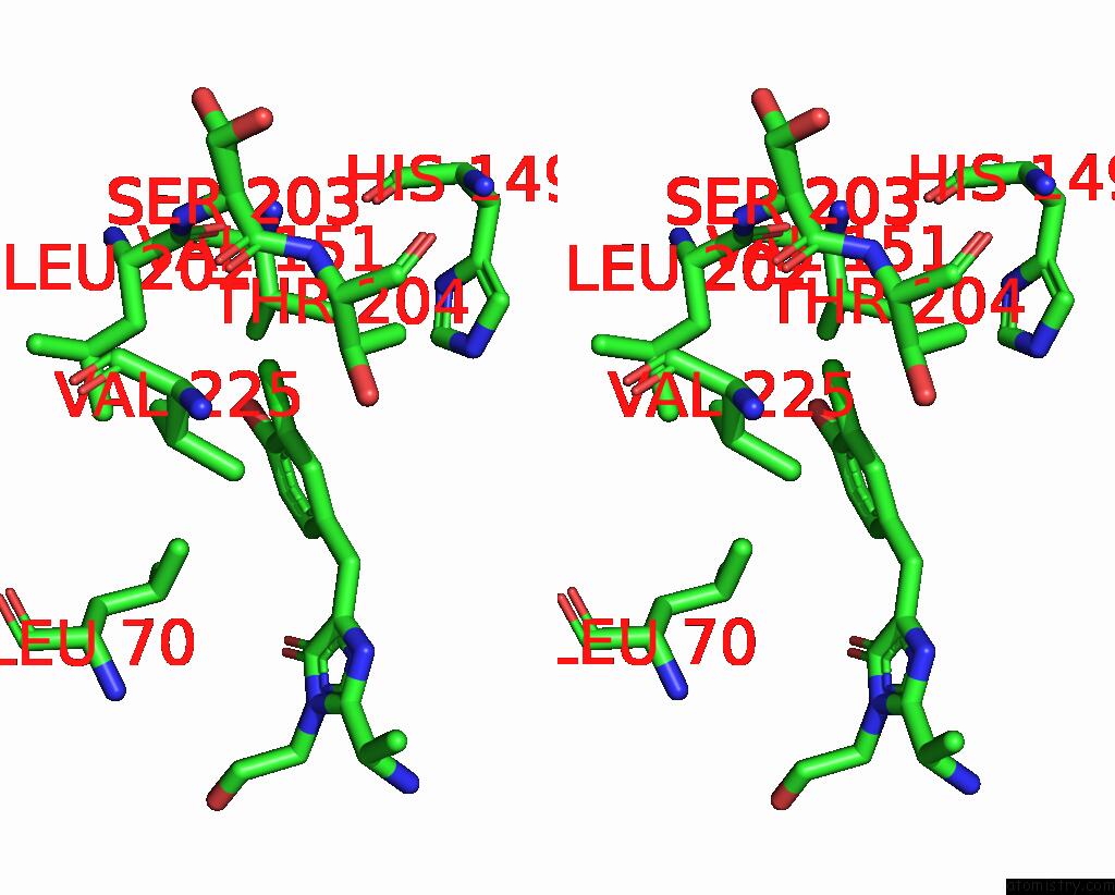
Stereo pair view
Mono view
Stereo pair view
A full contact list of Chlorine with other atoms in the Cl binding
site number 1 of RSEGFP2 with A Chlorinated Chromophore in the Non-Fluorescent Off- State within 5.0Å range:
|
Reference:
J.Chang,
M.G.Romei,
S.G.Boxer.
Structural Evidence of Photoisomerization Pathways in Fluorescent Proteins. J.Am.Chem.Soc. V. 141 15504 2019.
ISSN: ESSN 1520-5126
PubMed: 31533429
DOI: 10.1021/JACS.9B08356
Page generated: Mon Jul 29 13:20:44 2024
ISSN: ESSN 1520-5126
PubMed: 31533429
DOI: 10.1021/JACS.9B08356
Last articles
Zn in 9J0NZn in 9J0O
Zn in 9J0P
Zn in 9FJX
Zn in 9EKB
Zn in 9C0F
Zn in 9CAH
Zn in 9CH0
Zn in 9CH3
Zn in 9CH1