Chlorine »
PDB 6prh-6q0m »
6pry »
Chlorine in PDB 6pry: X-Ray Crystal Structure of the Blue-Light Absorbing State of Pixj From Thermosynechococcus Elongatus By Serial Femtosecond Crystallographic Analysis
Protein crystallography data
The structure of X-Ray Crystal Structure of the Blue-Light Absorbing State of Pixj From Thermosynechococcus Elongatus By Serial Femtosecond Crystallographic Analysis, PDB code: 6pry
was solved by
E.S.Burgie,
J.A.Clinger,
M.D.Miller,
G.N.Phillips Jr.,
R.D.Vierstra,
A.M.Orville,
J.F.Kern,
with X-Ray Crystallography technique. A brief refinement statistics is given in the table below:
| Resolution Low / High (Å) | 24.09 / 1.55 |
| Space group | P 21 21 21 |
| Cell size a, b, c (Å), α, β, γ (°) | 44.788, 61.417, 116.484, 90.00, 90.00, 90.00 |
| R / Rfree (%) | 15.7 / 18.5 |
Other elements in 6pry:
The structure of X-Ray Crystal Structure of the Blue-Light Absorbing State of Pixj From Thermosynechococcus Elongatus By Serial Femtosecond Crystallographic Analysis also contains other interesting chemical elements:
| Magnesium | (Mg) | 4 atoms |
Chlorine Binding Sites:
The binding sites of Chlorine atom in the X-Ray Crystal Structure of the Blue-Light Absorbing State of Pixj From Thermosynechococcus Elongatus By Serial Femtosecond Crystallographic Analysis
(pdb code 6pry). This binding sites where shown within
5.0 Angstroms radius around Chlorine atom.
In total 3 binding sites of Chlorine where determined in the X-Ray Crystal Structure of the Blue-Light Absorbing State of Pixj From Thermosynechococcus Elongatus By Serial Femtosecond Crystallographic Analysis, PDB code: 6pry:
Jump to Chlorine binding site number: 1; 2; 3;
In total 3 binding sites of Chlorine where determined in the X-Ray Crystal Structure of the Blue-Light Absorbing State of Pixj From Thermosynechococcus Elongatus By Serial Femtosecond Crystallographic Analysis, PDB code: 6pry:
Jump to Chlorine binding site number: 1; 2; 3;
Chlorine binding site 1 out of 3 in 6pry
Go back to
Chlorine binding site 1 out
of 3 in the X-Ray Crystal Structure of the Blue-Light Absorbing State of Pixj From Thermosynechococcus Elongatus By Serial Femtosecond Crystallographic Analysis
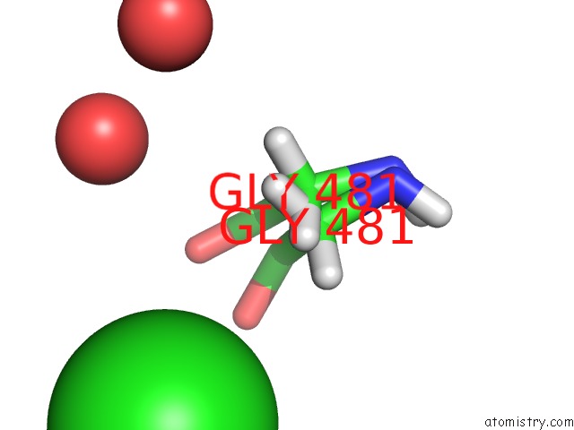
Mono view

Stereo pair view
Mono view
Stereo pair view
A full contact list of Chlorine with other atoms in the Cl binding
site number 1 of X-Ray Crystal Structure of the Blue-Light Absorbing State of Pixj From Thermosynechococcus Elongatus By Serial Femtosecond Crystallographic Analysis within 5.0Å range:
|
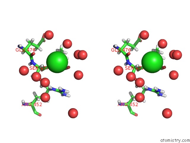
Chlorine binding site 2 out of 3 in 6pry
Go back to
Chlorine binding site 2 out
of 3 in the X-Ray Crystal Structure of the Blue-Light Absorbing State of Pixj From Thermosynechococcus Elongatus By Serial Femtosecond Crystallographic Analysis
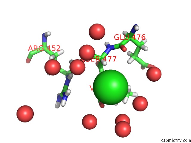
Mono view
Stereo pair view
Mono view
Stereo pair view
A full contact list of Chlorine with other atoms in the Cl binding
site number 2 of X-Ray Crystal Structure of the Blue-Light Absorbing State of Pixj From Thermosynechococcus Elongatus By Serial Femtosecond Crystallographic Analysis within 5.0Å range:
|
Chlorine binding site 3 out of 3 in 6pry
Go back to
Chlorine binding site 3 out
of 3 in the X-Ray Crystal Structure of the Blue-Light Absorbing State of Pixj From Thermosynechococcus Elongatus By Serial Femtosecond Crystallographic Analysis

Mono view

Stereo pair view
Mono view
Stereo pair view
A full contact list of Chlorine with other atoms in the Cl binding
site number 3 of X-Ray Crystal Structure of the Blue-Light Absorbing State of Pixj From Thermosynechococcus Elongatus By Serial Femtosecond Crystallographic Analysis within 5.0Å range:
|
Reference:
E.S.Burgie,
J.A.Clinger,
M.D.Miller,
A.S.Brewster,
P.Aller,
A.Butryn,
F.D.Fuller,
S.Gul,
I.D.Young,
C.C.Pham,
I.S.Kim,
A.Bhowmick,
L.J.O'riordan,
K.D.Sutherlin,
J.V.Heinemann,
A.Batyuk,
R.Alonso-Mori,
M.S.Hunter,
J.E.Koglin,
J.Yano,
V.K.Yachandra,
N.K.Sauter,
A.E.Cohen,
J.Kern,
A.M.Orville,
G.N.Phillips Jr.,
R.D.Vierstra.
Photoreversible Interconversion of A Phytochrome Photosensory Module in the Crystalline State. Proc.Natl.Acad.Sci.Usa 2019.
ISSN: ESSN 1091-6490
PubMed: 31852825
DOI: 10.1073/PNAS.1912041116
Page generated: Mon Jul 29 13:34:08 2024
ISSN: ESSN 1091-6490
PubMed: 31852825
DOI: 10.1073/PNAS.1912041116
Last articles
Zn in 9J0NZn in 9J0O
Zn in 9J0P
Zn in 9FJX
Zn in 9EKB
Zn in 9C0F
Zn in 9CAH
Zn in 9CH0
Zn in 9CH3
Zn in 9CH1