Chlorine »
PDB 6q0q-6q91 »
6q2p »
Chlorine in PDB 6q2p: Crystal Structure of Mouse Viperin Bound to Cytidine Triphosphate and S-Adenosylhomocysteine
Protein crystallography data
The structure of Crystal Structure of Mouse Viperin Bound to Cytidine Triphosphate and S-Adenosylhomocysteine, PDB code: 6q2p
was solved by
M.K.Fenwick,
M.Dong,
H.Lin,
S.E.Ealick,
with X-Ray Crystallography technique. A brief refinement statistics is given in the table below:
| Resolution Low / High (Å) | 39.77 / 1.45 |
| Space group | P 21 21 21 |
| Cell size a, b, c (Å), α, β, γ (°) | 36.483, 142.588, 143.732, 90.00, 90.00, 90.00 |
| R / Rfree (%) | 14.1 / 16 |
Other elements in 6q2p:
The structure of Crystal Structure of Mouse Viperin Bound to Cytidine Triphosphate and S-Adenosylhomocysteine also contains other interesting chemical elements:
| Iron | (Fe) | 8 atoms |
Chlorine Binding Sites:
The binding sites of Chlorine atom in the Crystal Structure of Mouse Viperin Bound to Cytidine Triphosphate and S-Adenosylhomocysteine
(pdb code 6q2p). This binding sites where shown within
5.0 Angstroms radius around Chlorine atom.
In total 3 binding sites of Chlorine where determined in the Crystal Structure of Mouse Viperin Bound to Cytidine Triphosphate and S-Adenosylhomocysteine, PDB code: 6q2p:
Jump to Chlorine binding site number: 1; 2; 3;
In total 3 binding sites of Chlorine where determined in the Crystal Structure of Mouse Viperin Bound to Cytidine Triphosphate and S-Adenosylhomocysteine, PDB code: 6q2p:
Jump to Chlorine binding site number: 1; 2; 3;
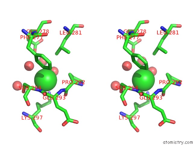
Chlorine binding site 1 out of 3 in 6q2p
Go back to
Chlorine binding site 1 out
of 3 in the Crystal Structure of Mouse Viperin Bound to Cytidine Triphosphate and S-Adenosylhomocysteine

Mono view

Stereo pair view
Mono view
Stereo pair view
A full contact list of Chlorine with other atoms in the Cl binding
site number 1 of Crystal Structure of Mouse Viperin Bound to Cytidine Triphosphate and S-Adenosylhomocysteine within 5.0Å range:
|
Chlorine binding site 2 out of 3 in 6q2p
Go back to
Chlorine binding site 2 out
of 3 in the Crystal Structure of Mouse Viperin Bound to Cytidine Triphosphate and S-Adenosylhomocysteine

Mono view

Stereo pair view
Mono view
Stereo pair view
A full contact list of Chlorine with other atoms in the Cl binding
site number 2 of Crystal Structure of Mouse Viperin Bound to Cytidine Triphosphate and S-Adenosylhomocysteine within 5.0Å range:
|
Chlorine binding site 3 out of 3 in 6q2p
Go back to
Chlorine binding site 3 out
of 3 in the Crystal Structure of Mouse Viperin Bound to Cytidine Triphosphate and S-Adenosylhomocysteine

Mono view
Stereo pair view
Mono view
Stereo pair view
A full contact list of Chlorine with other atoms in the Cl binding
site number 3 of Crystal Structure of Mouse Viperin Bound to Cytidine Triphosphate and S-Adenosylhomocysteine within 5.0Å range:
|
Reference:
M.K.Fenwick,
D.Su,
M.Dong,
H.Lin,
S.E.Ealick.
Structural Basis of Substrate Selectivity of Viperin. Biochemistry 2020.
ISSN: ISSN 0006-2960
PubMed: 31917549
DOI: 10.1021/ACS.BIOCHEM.9B00741
Page generated: Mon Jul 29 13:42:47 2024
ISSN: ISSN 0006-2960
PubMed: 31917549
DOI: 10.1021/ACS.BIOCHEM.9B00741
Last articles
Zn in 9J0NZn in 9J0O
Zn in 9J0P
Zn in 9FJX
Zn in 9EKB
Zn in 9C0F
Zn in 9CAH
Zn in 9CH0
Zn in 9CH3
Zn in 9CH1