Chlorine »
PDB 6q96-6qig »
6qdr »
Chlorine in PDB 6qdr: Crystal Structure of 14-3-3SIGMA in Complex with A PAK6 PT99 Phosphopeptide
Enzymatic activity of Crystal Structure of 14-3-3SIGMA in Complex with A PAK6 PT99 Phosphopeptide
All present enzymatic activity of Crystal Structure of 14-3-3SIGMA in Complex with A PAK6 PT99 Phosphopeptide:
2.7.11.1;
2.7.11.1;
Protein crystallography data
The structure of Crystal Structure of 14-3-3SIGMA in Complex with A PAK6 PT99 Phosphopeptide, PDB code: 6qdr
was solved by
S.A.Andrei,
A.Kaplan,
A.E.Fournier,
C.Ottman,
with X-Ray Crystallography technique. A brief refinement statistics is given in the table below:
| Resolution Low / High (Å) | 41.84 / 1.62 |
| Space group | C 2 2 21 |
| Cell size a, b, c (Å), α, β, γ (°) | 82.479, 112.397, 62.667, 90.00, 90.00, 90.00 |
| R / Rfree (%) | 15.1 / 18.2 |
Other elements in 6qdr:
The structure of Crystal Structure of 14-3-3SIGMA in Complex with A PAK6 PT99 Phosphopeptide also contains other interesting chemical elements:
| Magnesium | (Mg) | 1 atom |
| Calcium | (Ca) | 2 atoms |
| Sodium | (Na) | 3 atoms |
Chlorine Binding Sites:
The binding sites of Chlorine atom in the Crystal Structure of 14-3-3SIGMA in Complex with A PAK6 PT99 Phosphopeptide
(pdb code 6qdr). This binding sites where shown within
5.0 Angstroms radius around Chlorine atom.
In total only one binding site of Chlorine was determined in the Crystal Structure of 14-3-3SIGMA in Complex with A PAK6 PT99 Phosphopeptide, PDB code: 6qdr:
In total only one binding site of Chlorine was determined in the Crystal Structure of 14-3-3SIGMA in Complex with A PAK6 PT99 Phosphopeptide, PDB code: 6qdr:
Chlorine binding site 1 out of 1 in 6qdr
Go back to
Chlorine binding site 1 out
of 1 in the Crystal Structure of 14-3-3SIGMA in Complex with A PAK6 PT99 Phosphopeptide
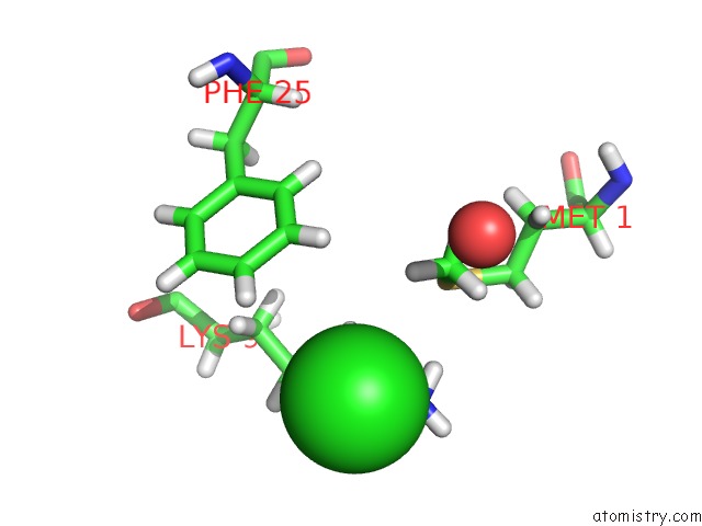
Mono view
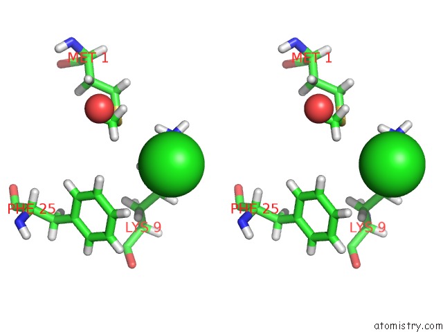
Stereo pair view
Mono view
Stereo pair view
A full contact list of Chlorine with other atoms in the Cl binding
site number 1 of Crystal Structure of 14-3-3SIGMA in Complex with A PAK6 PT99 Phosphopeptide within 5.0Å range:
|
Reference:
A.Kaplan,
S.A.Andrei,
C.Ottmann,
A.E.Fournier.
A Single-Agent Polypharmacological Approach to Stimulate Axon Regeneration To Be Published.
Page generated: Mon Jul 29 13:54:39 2024
Last articles
Ca in 5QJ3Ca in 5PTP
Ca in 5PB6
Ca in 5PB5
Ca in 5PB4
Ca in 5PB3
Ca in 5PB2
Ca in 5PB0
Ca in 5PB1
Ca in 5PAY