Chlorine »
PDB 6q96-6qig »
6qgi »
Chlorine in PDB 6qgi: Crystal Structure of VP5 From Haloarchaeal Pleomorphic Virus 2
Protein crystallography data
The structure of Crystal Structure of VP5 From Haloarchaeal Pleomorphic Virus 2, PDB code: 6qgi
was solved by
K.El Omari,
T.S.Walter,
K.Harlos,
J.M.Grimes,
D.I.Stuart,
E.Roine,
with X-Ray Crystallography technique. A brief refinement statistics is given in the table below:
| Resolution Low / High (Å) | 46.60 / 2.46 |
| Space group | P 21 21 21 |
| Cell size a, b, c (Å), α, β, γ (°) | 48.080, 93.220, 121.820, 90.00, 90.00, 90.00 |
| R / Rfree (%) | 22.4 / 24.1 |
Chlorine Binding Sites:
The binding sites of Chlorine atom in the Crystal Structure of VP5 From Haloarchaeal Pleomorphic Virus 2
(pdb code 6qgi). This binding sites where shown within
5.0 Angstroms radius around Chlorine atom.
In total 4 binding sites of Chlorine where determined in the Crystal Structure of VP5 From Haloarchaeal Pleomorphic Virus 2, PDB code: 6qgi:
Jump to Chlorine binding site number: 1; 2; 3; 4;
In total 4 binding sites of Chlorine where determined in the Crystal Structure of VP5 From Haloarchaeal Pleomorphic Virus 2, PDB code: 6qgi:
Jump to Chlorine binding site number: 1; 2; 3; 4;
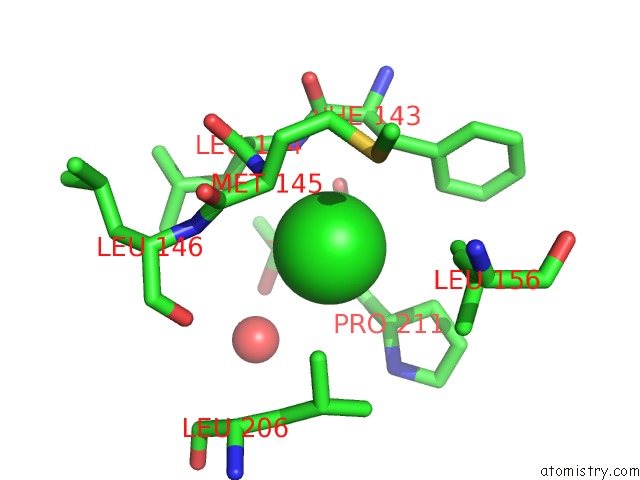
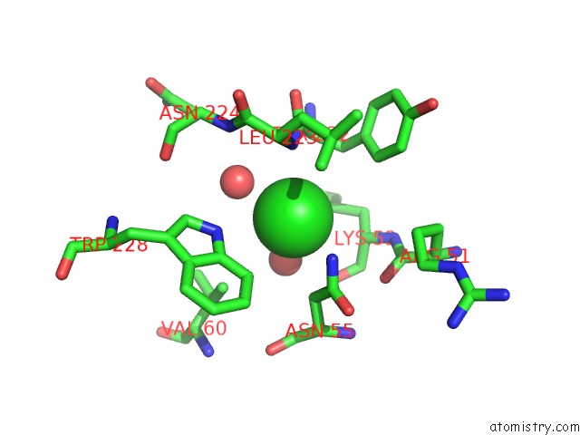
Chlorine binding site 1 out of 4 in 6qgi
Go back to
Chlorine binding site 1 out
of 4 in the Crystal Structure of VP5 From Haloarchaeal Pleomorphic Virus 2
Mono view
Stereo pair view
Mono view
Stereo pair view
A full contact list of Chlorine with other atoms in the Cl binding
site number 1 of Crystal Structure of VP5 From Haloarchaeal Pleomorphic Virus 2 within 5.0Å range:
|
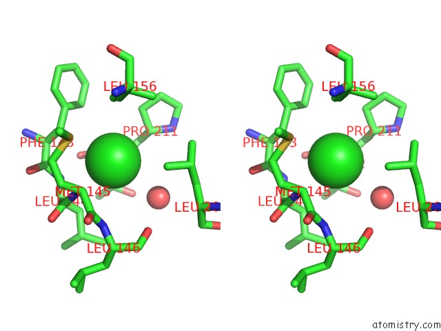
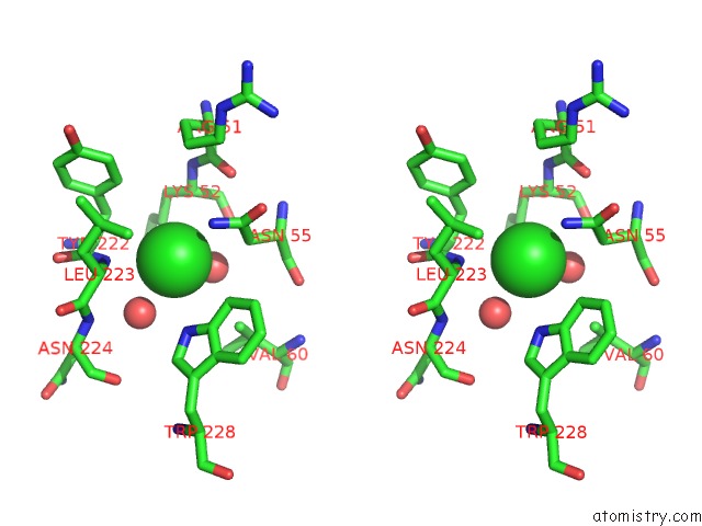
Chlorine binding site 2 out of 4 in 6qgi
Go back to
Chlorine binding site 2 out
of 4 in the Crystal Structure of VP5 From Haloarchaeal Pleomorphic Virus 2
Mono view
Stereo pair view
Mono view
Stereo pair view
A full contact list of Chlorine with other atoms in the Cl binding
site number 2 of Crystal Structure of VP5 From Haloarchaeal Pleomorphic Virus 2 within 5.0Å range:
|
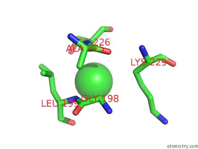
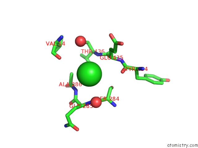
Chlorine binding site 3 out of 4 in 6qgi
Go back to
Chlorine binding site 3 out
of 4 in the Crystal Structure of VP5 From Haloarchaeal Pleomorphic Virus 2
Mono view
Stereo pair view
Mono view
Stereo pair view
A full contact list of Chlorine with other atoms in the Cl binding
site number 3 of Crystal Structure of VP5 From Haloarchaeal Pleomorphic Virus 2 within 5.0Å range:
|
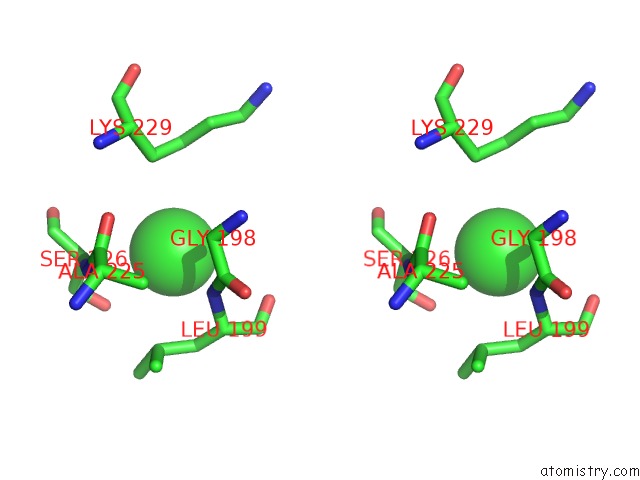
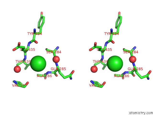
Chlorine binding site 4 out of 4 in 6qgi
Go back to
Chlorine binding site 4 out
of 4 in the Crystal Structure of VP5 From Haloarchaeal Pleomorphic Virus 2
Mono view
Stereo pair view
Mono view
Stereo pair view
A full contact list of Chlorine with other atoms in the Cl binding
site number 4 of Crystal Structure of VP5 From Haloarchaeal Pleomorphic Virus 2 within 5.0Å range:
|
Reference:
K.El Omari,
S.Li,
A.Kotecha,
T.S.Walter,
E.A.Bignon,
K.Harlos,
P.Somerharju,
F.De Haas,
D.K.Clare,
M.Molin,
F.Hurtado,
M.Li,
J.M.Grimes,
D.H.Bamford,
N.D.Tischler,
J.T.Huiskonen,
D.I.Stuart,
E.Roine.
The Structure of A Prokaryotic Viral Envelope Protein Expands the Landscape of Membrane Fusion Proteins. Nat Commun V. 10 846 2019.
ISSN: ESSN 2041-1723
PubMed: 30783086
DOI: 10.1038/S41467-019-08728-7
Page generated: Mon Jul 29 13:57:05 2024
ISSN: ESSN 2041-1723
PubMed: 30783086
DOI: 10.1038/S41467-019-08728-7
Last articles
Zn in 9J0NZn in 9J0O
Zn in 9J0P
Zn in 9FJX
Zn in 9EKB
Zn in 9C0F
Zn in 9CAH
Zn in 9CH0
Zn in 9CH3
Zn in 9CH1