Chlorine »
PDB 6ris-6ro5 »
6rjn »
Chlorine in PDB 6rjn: Crystal Structure of A Fungal Catalase at 2.3 Angstroms
Enzymatic activity of Crystal Structure of A Fungal Catalase at 2.3 Angstroms
All present enzymatic activity of Crystal Structure of A Fungal Catalase at 2.3 Angstroms:
1.11.1.6;
1.11.1.6;
Protein crystallography data
The structure of Crystal Structure of A Fungal Catalase at 2.3 Angstroms, PDB code: 6rjn
was solved by
S.Gomez,
S.Navas-Yuste,
A.M.Payne,
W.Rivera,
M.Lopez-Estepa,
C.Brangbour,
D.Fulla,
J.Juanhuix,
F.J.Fernandez,
M.C.Vega,
with X-Ray Crystallography technique. A brief refinement statistics is given in the table below:
| Resolution Low / High (Å) | 54.67 / 2.30 |
| Space group | P 21 21 2 |
| Cell size a, b, c (Å), α, β, γ (°) | 165.943, 173.690, 96.490, 90.00, 90.00, 90.00 |
| R / Rfree (%) | 14.3 / 19.6 |
Other elements in 6rjn:
The structure of Crystal Structure of A Fungal Catalase at 2.3 Angstroms also contains other interesting chemical elements:
| Potassium | (K) | 7 atoms |
| Iron | (Fe) | 4 atoms |
| Sodium | (Na) | 4 atoms |
Chlorine Binding Sites:
The binding sites of Chlorine atom in the Crystal Structure of A Fungal Catalase at 2.3 Angstroms
(pdb code 6rjn). This binding sites where shown within
5.0 Angstroms radius around Chlorine atom.
In total 5 binding sites of Chlorine where determined in the Crystal Structure of A Fungal Catalase at 2.3 Angstroms, PDB code: 6rjn:
Jump to Chlorine binding site number: 1; 2; 3; 4; 5;
In total 5 binding sites of Chlorine where determined in the Crystal Structure of A Fungal Catalase at 2.3 Angstroms, PDB code: 6rjn:
Jump to Chlorine binding site number: 1; 2; 3; 4; 5;
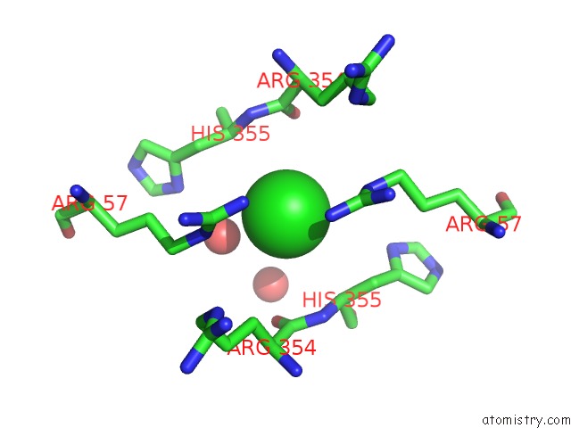
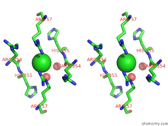
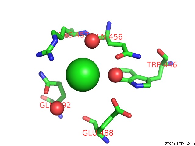
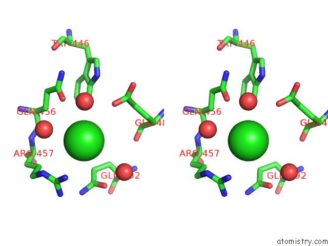
Chlorine binding site 1 out of 5 in 6rjn
Go back to
Chlorine binding site 1 out
of 5 in the Crystal Structure of A Fungal Catalase at 2.3 Angstroms
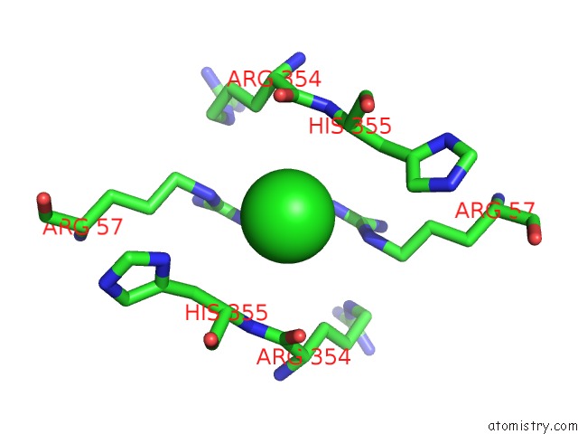
Mono view
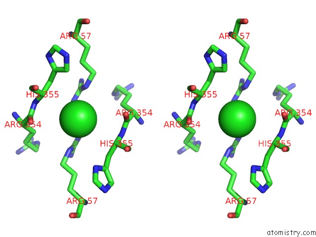
Stereo pair view
Mono view
Stereo pair view
A full contact list of Chlorine with other atoms in the Cl binding
site number 1 of Crystal Structure of A Fungal Catalase at 2.3 Angstroms within 5.0Å range:
|
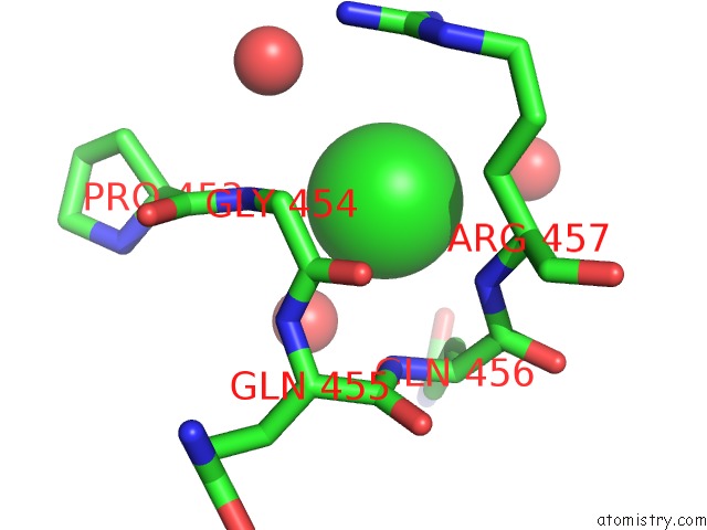
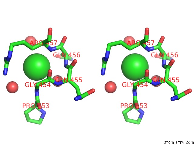
Chlorine binding site 2 out of 5 in 6rjn
Go back to
Chlorine binding site 2 out
of 5 in the Crystal Structure of A Fungal Catalase at 2.3 Angstroms
Mono view
Stereo pair view
Mono view
Stereo pair view
A full contact list of Chlorine with other atoms in the Cl binding
site number 2 of Crystal Structure of A Fungal Catalase at 2.3 Angstroms within 5.0Å range:
|
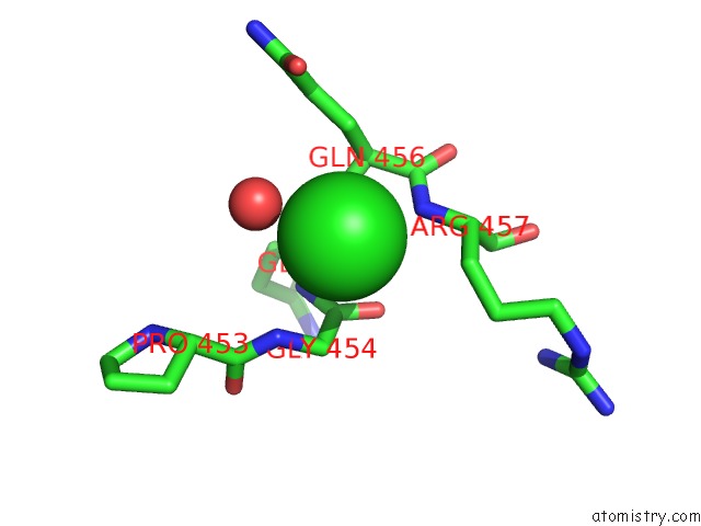
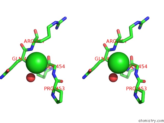
Chlorine binding site 3 out of 5 in 6rjn
Go back to
Chlorine binding site 3 out
of 5 in the Crystal Structure of A Fungal Catalase at 2.3 Angstroms
Mono view
Stereo pair view
Mono view
Stereo pair view
A full contact list of Chlorine with other atoms in the Cl binding
site number 3 of Crystal Structure of A Fungal Catalase at 2.3 Angstroms within 5.0Å range:
|
Chlorine binding site 4 out of 5 in 6rjn
Go back to
Chlorine binding site 4 out
of 5 in the Crystal Structure of A Fungal Catalase at 2.3 Angstroms
Mono view
Stereo pair view
Mono view
Stereo pair view
A full contact list of Chlorine with other atoms in the Cl binding
site number 4 of Crystal Structure of A Fungal Catalase at 2.3 Angstroms within 5.0Å range:
|
Chlorine binding site 5 out of 5 in 6rjn
Go back to
Chlorine binding site 5 out
of 5 in the Crystal Structure of A Fungal Catalase at 2.3 Angstroms
Mono view
Stereo pair view
Mono view
Stereo pair view
A full contact list of Chlorine with other atoms in the Cl binding
site number 5 of Crystal Structure of A Fungal Catalase at 2.3 Angstroms within 5.0Å range:
|
Reference:
S.Gomez,
S.Navas-Yuste,
A.M.Payne,
W.Rivera,
M.Lopez-Estepa,
C.Brangbour,
D.Fulla,
J.Juanhuix,
F.J.Fernandez,
M.C.Vega.
Peroxisomal Catalases From the Yeasts Pichia Pastoris and Kluyveromyces Lactis As Models For Oxidative Damage in Higher Eukaryotes. Free Radic. Biol. Med. V. 141 279 2019.
ISSN: ISSN 1873-4596
PubMed: 31238127
DOI: 10.1016/J.FREERADBIOMED.2019.06.025
Page generated: Mon Jul 29 14:31:53 2024
ISSN: ISSN 1873-4596
PubMed: 31238127
DOI: 10.1016/J.FREERADBIOMED.2019.06.025
Last articles
Cl in 5U27Cl in 5U2K
Cl in 5U2A
Cl in 5U24
Cl in 5U23
Cl in 5U21
Cl in 5U1R
Cl in 5U1A
Cl in 5U1Z
Cl in 5U20