Chlorine »
PDB 6ris-6ro5 »
6rlm »
Chlorine in PDB 6rlm: Crystal Structure of AT1412DM Fab Fragment
Protein crystallography data
The structure of Crystal Structure of AT1412DM Fab Fragment, PDB code: 6rlm
was solved by
V.Neviani,
N.M.Pearce,
W.Pos,
R.Schotte,
H.Spits,
P.Gros,
with X-Ray Crystallography technique. A brief refinement statistics is given in the table below:
| Resolution Low / High (Å) | 163.66 / 2.50 |
| Space group | C 2 2 21 |
| Cell size a, b, c (Å), α, β, γ (°) | 224.909, 238.594, 209.812, 90, 90, 90 |
| R / Rfree (%) | 24.2 / 26.7 |
Chlorine Binding Sites:
The binding sites of Chlorine atom in the Crystal Structure of AT1412DM Fab Fragment
(pdb code 6rlm). This binding sites where shown within
5.0 Angstroms radius around Chlorine atom.
In total 4 binding sites of Chlorine where determined in the Crystal Structure of AT1412DM Fab Fragment, PDB code: 6rlm:
Jump to Chlorine binding site number: 1; 2; 3; 4;
In total 4 binding sites of Chlorine where determined in the Crystal Structure of AT1412DM Fab Fragment, PDB code: 6rlm:
Jump to Chlorine binding site number: 1; 2; 3; 4;
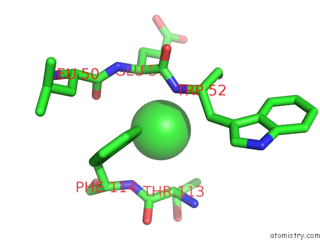
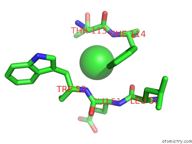
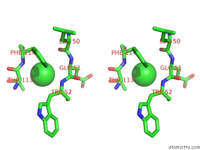
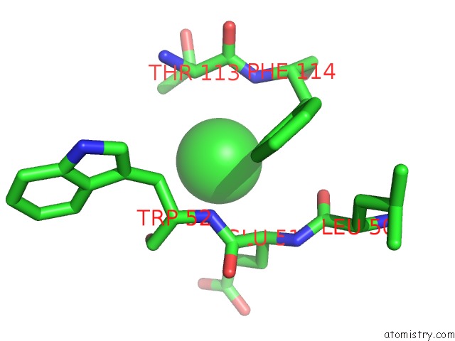
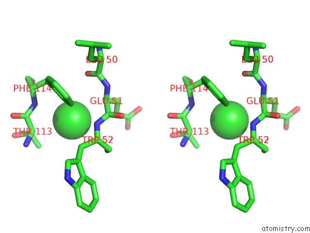
Chlorine binding site 1 out of 4 in 6rlm
Go back to
Chlorine binding site 1 out
of 4 in the Crystal Structure of AT1412DM Fab Fragment
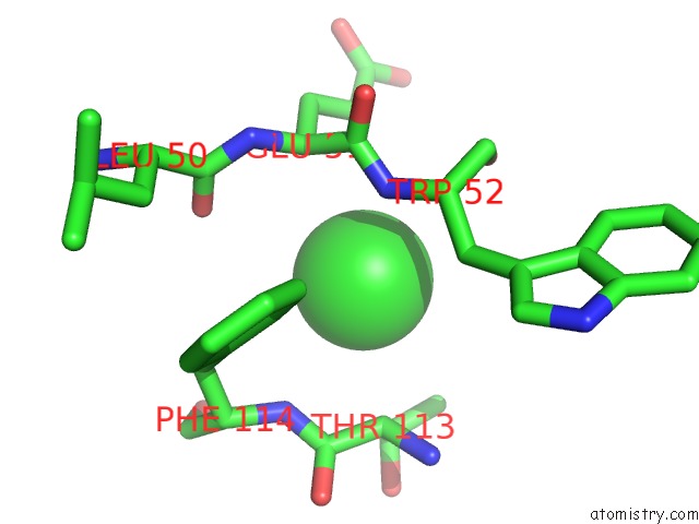
Mono view
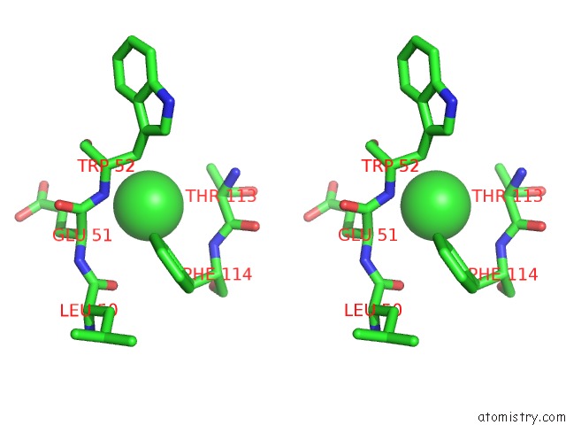
Stereo pair view
Mono view
Stereo pair view
A full contact list of Chlorine with other atoms in the Cl binding
site number 1 of Crystal Structure of AT1412DM Fab Fragment within 5.0Å range:
|
Chlorine binding site 2 out of 4 in 6rlm
Go back to
Chlorine binding site 2 out
of 4 in the Crystal Structure of AT1412DM Fab Fragment
Mono view

Stereo pair view
Mono view
Stereo pair view
A full contact list of Chlorine with other atoms in the Cl binding
site number 2 of Crystal Structure of AT1412DM Fab Fragment within 5.0Å range:
|
Chlorine binding site 3 out of 4 in 6rlm
Go back to
Chlorine binding site 3 out
of 4 in the Crystal Structure of AT1412DM Fab Fragment
Mono view
Stereo pair view
Mono view
Stereo pair view
A full contact list of Chlorine with other atoms in the Cl binding
site number 3 of Crystal Structure of AT1412DM Fab Fragment within 5.0Å range:
|
Chlorine binding site 4 out of 4 in 6rlm
Go back to
Chlorine binding site 4 out
of 4 in the Crystal Structure of AT1412DM Fab Fragment
Mono view
Stereo pair view
Mono view
Stereo pair view
A full contact list of Chlorine with other atoms in the Cl binding
site number 4 of Crystal Structure of AT1412DM Fab Fragment within 5.0Å range:
|
Reference:
V.Neviani,
W.Pos,
R.Schotte,
K.Wagner,
D.M.Go,
C.Fatmawati,
M.Kedde,
Y.B.Claassen,
L.Kroon-Batenburg,
M.Lutz,
E.M.E.Verdegaal,
S.H.Van Der Burg,
H.Spits,
P.Gros.
Structural Basis of A Homo-Dimerization Site in Tetraspanin CD9 Targeted By A Melanoma Patient-Derived Antibody To Be Published.
Page generated: Mon Jul 29 14:33:56 2024
Last articles
Zn in 9J0NZn in 9J0O
Zn in 9J0P
Zn in 9FJX
Zn in 9EKB
Zn in 9C0F
Zn in 9CAH
Zn in 9CH0
Zn in 9CH3
Zn in 9CH1