Chlorine »
PDB 6rot-6rzp »
6rs6 »
Chlorine in PDB 6rs6: X-Ray Crystal Structure of LSAA9B
Protein crystallography data
The structure of X-Ray Crystal Structure of LSAA9B, PDB code: 6rs6
was solved by
K.E.H.Frandsen,
M.Tovborg,
J.C.N.Poulsen,
K.S.Johansen,
L.Lo Leggio,
with X-Ray Crystallography technique. A brief refinement statistics is given in the table below:
| Resolution Low / High (Å) | 19.93 / 1.60 |
| Space group | P 21 21 21 |
| Cell size a, b, c (Å), α, β, γ (°) | 35.190, 72.480, 78.560, 90.00, 90.00, 90.00 |
| R / Rfree (%) | 13.7 / 18.2 |
Chlorine Binding Sites:
The binding sites of Chlorine atom in the X-Ray Crystal Structure of LSAA9B
(pdb code 6rs6). This binding sites where shown within
5.0 Angstroms radius around Chlorine atom.
In total 2 binding sites of Chlorine where determined in the X-Ray Crystal Structure of LSAA9B, PDB code: 6rs6:
Jump to Chlorine binding site number: 1; 2;
In total 2 binding sites of Chlorine where determined in the X-Ray Crystal Structure of LSAA9B, PDB code: 6rs6:
Jump to Chlorine binding site number: 1; 2;
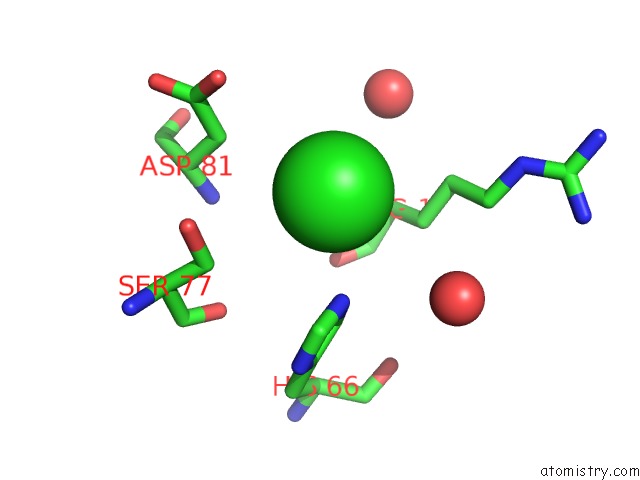
Chlorine binding site 1 out of 2 in 6rs6
Go back to
Chlorine binding site 1 out
of 2 in the X-Ray Crystal Structure of LSAA9B
Mono view

Stereo pair view
Mono view
Stereo pair view
A full contact list of Chlorine with other atoms in the Cl binding
site number 1 of X-Ray Crystal Structure of LSAA9B within 5.0Å range:
|
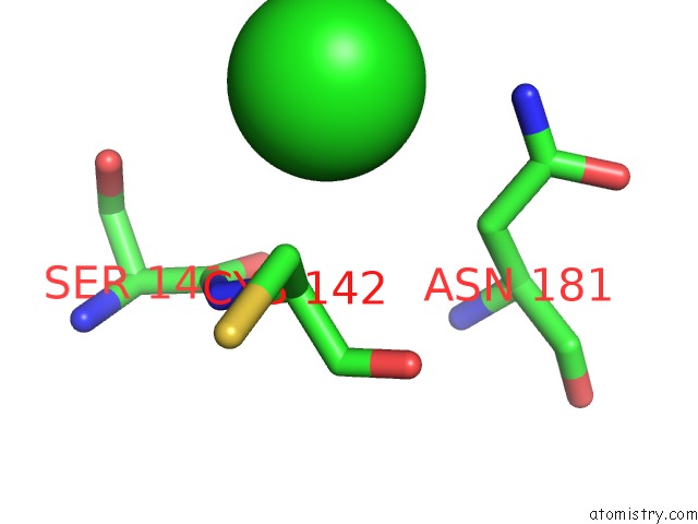
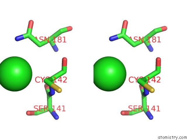
Chlorine binding site 2 out of 2 in 6rs6
Go back to
Chlorine binding site 2 out
of 2 in the X-Ray Crystal Structure of LSAA9B
Mono view
Stereo pair view
Mono view
Stereo pair view
A full contact list of Chlorine with other atoms in the Cl binding
site number 2 of X-Ray Crystal Structure of LSAA9B within 5.0Å range:
|
Reference:
K.E.H.Frandsen,
M.Tovborg,
C.I.Jorgensen,
N.Spodsberg,
M.N.Rosso,
G.R.Hemsworth,
E.F.Garman,
G.W.Grime,
J.N.Poulsen,
T.S.Batth,
S.Miyauchi,
A.Lipzen,
C.Daum,
I.V.Grigoriev,
K.S.Johansen,
B.Henrissat,
J.G.Berrin,
L.Lo Leggio.
Insights Into An Unusual Auxiliary Activity 9 Family Member Lacking the Histidine Brace Motif of Lytic Polysaccharide Monooxygenases. J.Biol.Chem. V. 294 17117 2019.
ISSN: ESSN 1083-351X
PubMed: 31471321
DOI: 10.1074/JBC.RA119.009223
Page generated: Mon Jul 29 14:38:34 2024
ISSN: ESSN 1083-351X
PubMed: 31471321
DOI: 10.1074/JBC.RA119.009223
Last articles
Zn in 9J0NZn in 9J0O
Zn in 9J0P
Zn in 9FJX
Zn in 9EKB
Zn in 9C0F
Zn in 9CAH
Zn in 9CH0
Zn in 9CH3
Zn in 9CH1