Chlorine »
PDB 6sa6-6sip »
6sbj »
Chlorine in PDB 6sbj: X-Ray Structure of Mus Musculus Fumarylacetoacetate Hydrolase Domain Containing Protein 1 (FAHD1) Apo-Form Uuncomplexed
Enzymatic activity of X-Ray Structure of Mus Musculus Fumarylacetoacetate Hydrolase Domain Containing Protein 1 (FAHD1) Apo-Form Uuncomplexed
All present enzymatic activity of X-Ray Structure of Mus Musculus Fumarylacetoacetate Hydrolase Domain Containing Protein 1 (FAHD1) Apo-Form Uuncomplexed:
3.7.1.5; 4.1.1.112;
3.7.1.5; 4.1.1.112;
Protein crystallography data
The structure of X-Ray Structure of Mus Musculus Fumarylacetoacetate Hydrolase Domain Containing Protein 1 (FAHD1) Apo-Form Uuncomplexed, PDB code: 6sbj
was solved by
B.Rupp,
A.Naschberger,
A.K.H.Weiss,
with X-Ray Crystallography technique. A brief refinement statistics is given in the table below:
| Resolution Low / High (Å) | 42.65 / 2.22 |
| Space group | P 1 21 1 |
| Cell size a, b, c (Å), α, β, γ (°) | 52.401, 69.959, 107.557, 90.00, 92.25, 90.00 |
| R / Rfree (%) | 19.3 / 25.7 |
Other elements in 6sbj:
The structure of X-Ray Structure of Mus Musculus Fumarylacetoacetate Hydrolase Domain Containing Protein 1 (FAHD1) Apo-Form Uuncomplexed also contains other interesting chemical elements:
| Magnesium | (Mg) | 6 atoms |
Chlorine Binding Sites:
The binding sites of Chlorine atom in the X-Ray Structure of Mus Musculus Fumarylacetoacetate Hydrolase Domain Containing Protein 1 (FAHD1) Apo-Form Uuncomplexed
(pdb code 6sbj). This binding sites where shown within
5.0 Angstroms radius around Chlorine atom.
In total 6 binding sites of Chlorine where determined in the X-Ray Structure of Mus Musculus Fumarylacetoacetate Hydrolase Domain Containing Protein 1 (FAHD1) Apo-Form Uuncomplexed, PDB code: 6sbj:
Jump to Chlorine binding site number: 1; 2; 3; 4; 5; 6;
In total 6 binding sites of Chlorine where determined in the X-Ray Structure of Mus Musculus Fumarylacetoacetate Hydrolase Domain Containing Protein 1 (FAHD1) Apo-Form Uuncomplexed, PDB code: 6sbj:
Jump to Chlorine binding site number: 1; 2; 3; 4; 5; 6;
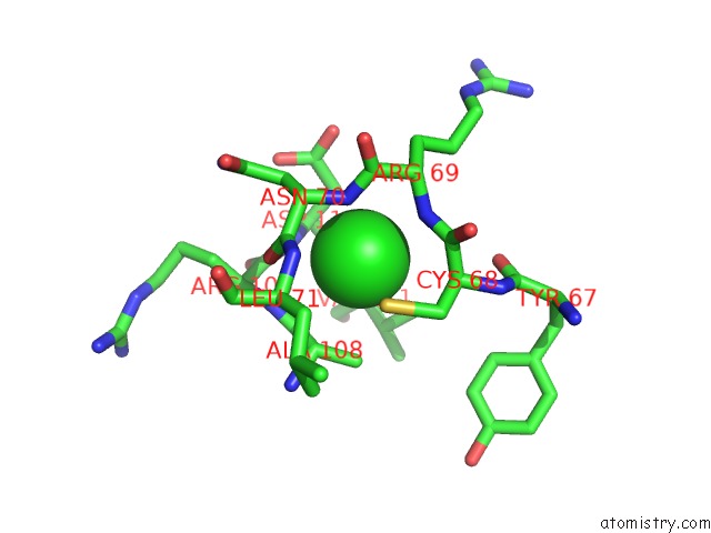
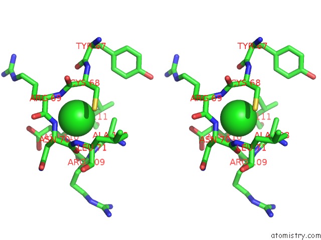
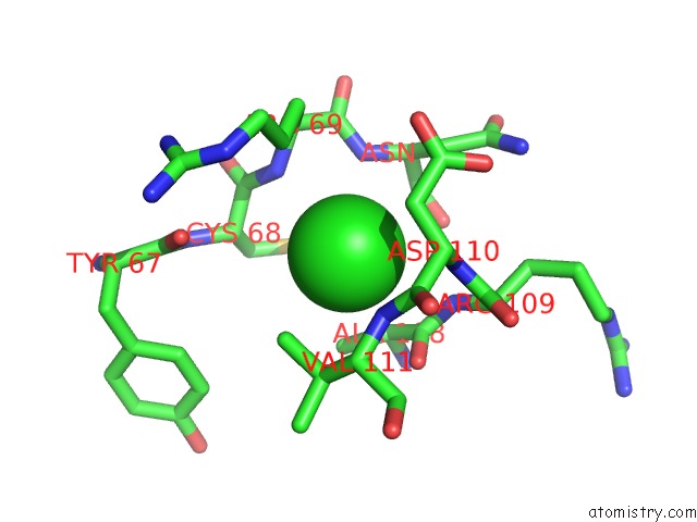
Chlorine binding site 1 out of 6 in 6sbj
Go back to
Chlorine binding site 1 out
of 6 in the X-Ray Structure of Mus Musculus Fumarylacetoacetate Hydrolase Domain Containing Protein 1 (FAHD1) Apo-Form Uuncomplexed
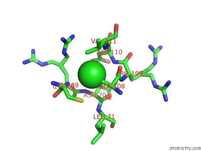
Mono view
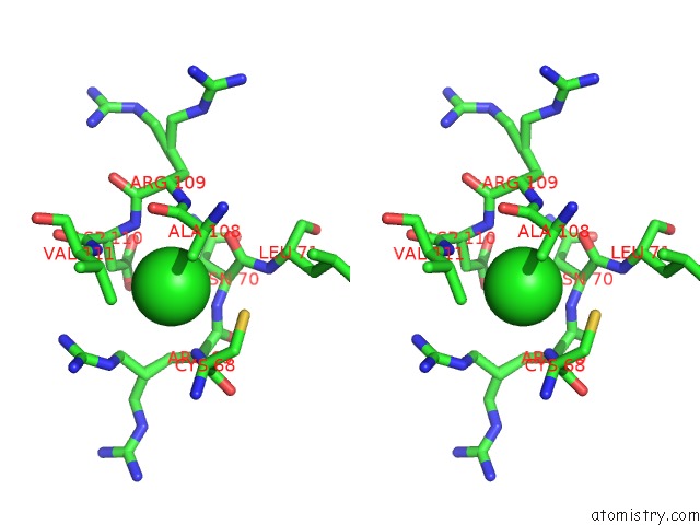
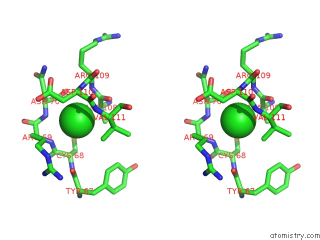
Stereo pair view
Mono view
Stereo pair view
A full contact list of Chlorine with other atoms in the Cl binding
site number 1 of X-Ray Structure of Mus Musculus Fumarylacetoacetate Hydrolase Domain Containing Protein 1 (FAHD1) Apo-Form Uuncomplexed within 5.0Å range:
|
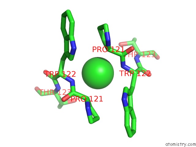
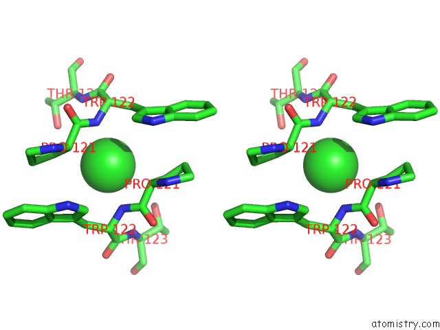
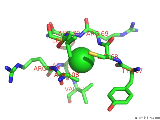
Chlorine binding site 2 out of 6 in 6sbj
Go back to
Chlorine binding site 2 out
of 6 in the X-Ray Structure of Mus Musculus Fumarylacetoacetate Hydrolase Domain Containing Protein 1 (FAHD1) Apo-Form Uuncomplexed
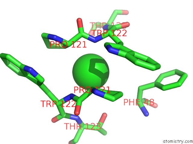
Mono view
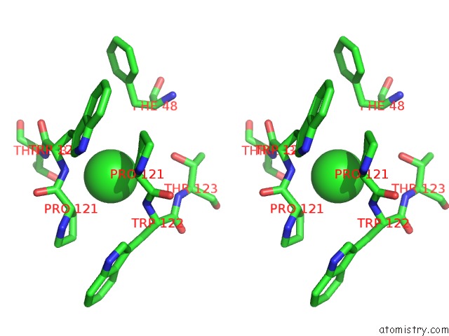
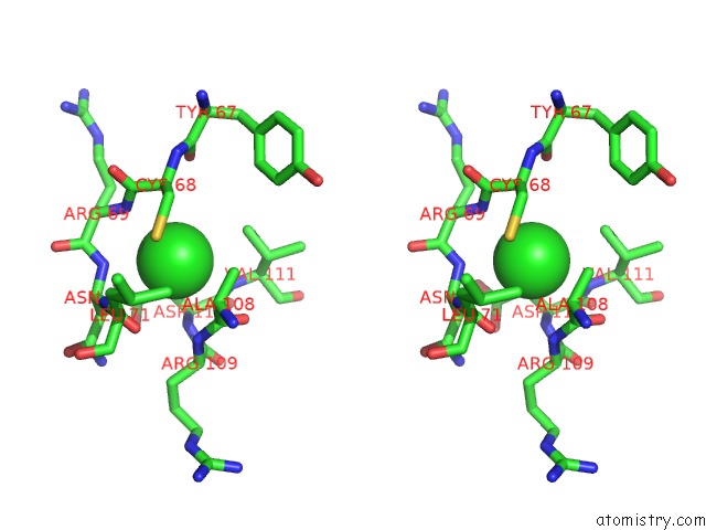
Stereo pair view
Mono view
Stereo pair view
A full contact list of Chlorine with other atoms in the Cl binding
site number 2 of X-Ray Structure of Mus Musculus Fumarylacetoacetate Hydrolase Domain Containing Protein 1 (FAHD1) Apo-Form Uuncomplexed within 5.0Å range:
|
Chlorine binding site 3 out of 6 in 6sbj
Go back to
Chlorine binding site 3 out
of 6 in the X-Ray Structure of Mus Musculus Fumarylacetoacetate Hydrolase Domain Containing Protein 1 (FAHD1) Apo-Form Uuncomplexed
Mono view
Stereo pair view
Mono view
Stereo pair view
A full contact list of Chlorine with other atoms in the Cl binding
site number 3 of X-Ray Structure of Mus Musculus Fumarylacetoacetate Hydrolase Domain Containing Protein 1 (FAHD1) Apo-Form Uuncomplexed within 5.0Å range:
|
Chlorine binding site 4 out of 6 in 6sbj
Go back to
Chlorine binding site 4 out
of 6 in the X-Ray Structure of Mus Musculus Fumarylacetoacetate Hydrolase Domain Containing Protein 1 (FAHD1) Apo-Form Uuncomplexed
Mono view
Stereo pair view
Mono view
Stereo pair view
A full contact list of Chlorine with other atoms in the Cl binding
site number 4 of X-Ray Structure of Mus Musculus Fumarylacetoacetate Hydrolase Domain Containing Protein 1 (FAHD1) Apo-Form Uuncomplexed within 5.0Å range:
|
Chlorine binding site 5 out of 6 in 6sbj
Go back to
Chlorine binding site 5 out
of 6 in the X-Ray Structure of Mus Musculus Fumarylacetoacetate Hydrolase Domain Containing Protein 1 (FAHD1) Apo-Form Uuncomplexed
Mono view
Stereo pair view
Mono view
Stereo pair view
A full contact list of Chlorine with other atoms in the Cl binding
site number 5 of X-Ray Structure of Mus Musculus Fumarylacetoacetate Hydrolase Domain Containing Protein 1 (FAHD1) Apo-Form Uuncomplexed within 5.0Å range:
|
Chlorine binding site 6 out of 6 in 6sbj
Go back to
Chlorine binding site 6 out
of 6 in the X-Ray Structure of Mus Musculus Fumarylacetoacetate Hydrolase Domain Containing Protein 1 (FAHD1) Apo-Form Uuncomplexed
Mono view
Stereo pair view
Mono view
Stereo pair view
A full contact list of Chlorine with other atoms in the Cl binding
site number 6 of X-Ray Structure of Mus Musculus Fumarylacetoacetate Hydrolase Domain Containing Protein 1 (FAHD1) Apo-Form Uuncomplexed within 5.0Å range:
|
Reference:
A.K.H.Weiss,
A.Naschberger,
E.Cappuccio,
C.Metzger,
L.Mottes,
M.Holzknecht,
J.Von Velsen,
M.W.Bowler,
B.Rupp,
P.Jansen-Durr.
Structural and Functional Comparison of Fumarylacetoacetate Domain Containing Protein 1 (FAHD1) in Human and Mouse. Biosci.Rep. 2020.
ISSN: ISSN 0144-8463
PubMed: 32068790
DOI: 10.1042/BSR20194431
Page generated: Mon Jul 29 14:52:46 2024
ISSN: ISSN 0144-8463
PubMed: 32068790
DOI: 10.1042/BSR20194431
Last articles
Zn in 9J0NZn in 9J0O
Zn in 9J0P
Zn in 9FJX
Zn in 9EKB
Zn in 9C0F
Zn in 9CAH
Zn in 9CH0
Zn in 9CH3
Zn in 9CH1