Chlorine »
PDB 6sa6-6sip »
6set »
Chlorine in PDB 6set: X-Ray Structure of the Gold/Lysozyme Adduct Formed Upon 3 Days Exposure of Protein Crystals to Compound 1
Enzymatic activity of X-Ray Structure of the Gold/Lysozyme Adduct Formed Upon 3 Days Exposure of Protein Crystals to Compound 1
All present enzymatic activity of X-Ray Structure of the Gold/Lysozyme Adduct Formed Upon 3 Days Exposure of Protein Crystals to Compound 1:
3.2.1.17;
3.2.1.17;
Protein crystallography data
The structure of X-Ray Structure of the Gold/Lysozyme Adduct Formed Upon 3 Days Exposure of Protein Crystals to Compound 1, PDB code: 6set
was solved by
G.Ferraro,
A.Giorgio,
A.Merlino,
with X-Ray Crystallography technique. A brief refinement statistics is given in the table below:
| Resolution Low / High (Å) | 30.86 / 1.90 |
| Space group | P 43 21 2 |
| Cell size a, b, c (Å), α, β, γ (°) | 77.448, 77.448, 37.316, 90.00, 90.00, 90.00 |
| R / Rfree (%) | 15.8 / 21.5 |
Other elements in 6set:
The structure of X-Ray Structure of the Gold/Lysozyme Adduct Formed Upon 3 Days Exposure of Protein Crystals to Compound 1 also contains other interesting chemical elements:
| Gold | (Au) | 5 atoms |
| Sodium | (Na) | 1 atom |
Chlorine Binding Sites:
The binding sites of Chlorine atom in the X-Ray Structure of the Gold/Lysozyme Adduct Formed Upon 3 Days Exposure of Protein Crystals to Compound 1
(pdb code 6set). This binding sites where shown within
5.0 Angstroms radius around Chlorine atom.
In total only one binding site of Chlorine was determined in the X-Ray Structure of the Gold/Lysozyme Adduct Formed Upon 3 Days Exposure of Protein Crystals to Compound 1, PDB code: 6set:
In total only one binding site of Chlorine was determined in the X-Ray Structure of the Gold/Lysozyme Adduct Formed Upon 3 Days Exposure of Protein Crystals to Compound 1, PDB code: 6set:
Chlorine binding site 1 out of 1 in 6set
Go back to
Chlorine binding site 1 out
of 1 in the X-Ray Structure of the Gold/Lysozyme Adduct Formed Upon 3 Days Exposure of Protein Crystals to Compound 1
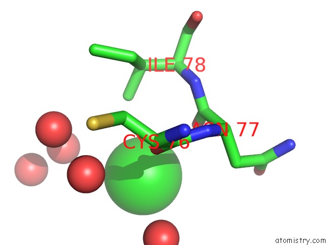
Mono view
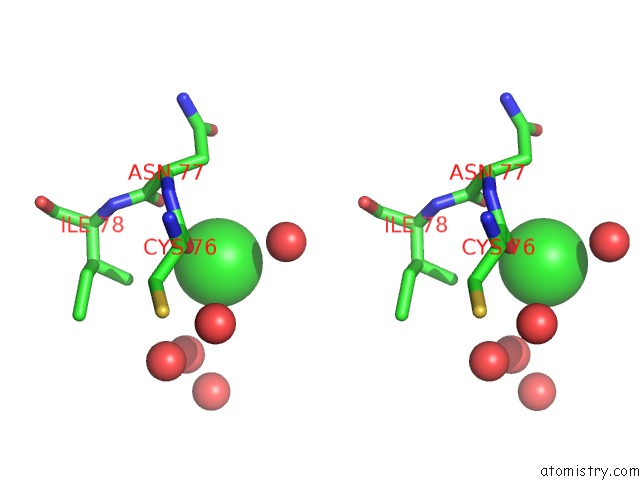
Stereo pair view
Mono view
Stereo pair view
A full contact list of Chlorine with other atoms in the Cl binding
site number 1 of X-Ray Structure of the Gold/Lysozyme Adduct Formed Upon 3 Days Exposure of Protein Crystals to Compound 1 within 5.0Å range:
|
Reference:
G.Ferraro,
A.Giorgio,
A.M.Mansour,
A.Merlino.
Protein-Mediated Disproportionation of Au(I): Insights From the Structures of Adducts of Au(III) Compounds Bearing N,N-Pyridylbenzimidazole Derivatives with Lysozyme. Dalton Trans V. 48 14027 2019.
ISSN: ESSN 1477-9234
PubMed: 31490509
DOI: 10.1039/C9DT02729G
Page generated: Mon Jul 29 14:56:51 2024
ISSN: ESSN 1477-9234
PubMed: 31490509
DOI: 10.1039/C9DT02729G
Last articles
Zn in 9MJ5Zn in 9HNW
Zn in 9G0L
Zn in 9FNE
Zn in 9DZN
Zn in 9E0I
Zn in 9D32
Zn in 9DAK
Zn in 8ZXC
Zn in 8ZUF