Chlorine »
PDB 6siq-6sr2 »
6sqb »
Chlorine in PDB 6sqb: Crystal Structure of M. Tuberculosis Inha in Complex with Nad+ and 3- (3-Chlorophenyl)Propanoic Acid
Enzymatic activity of Crystal Structure of M. Tuberculosis Inha in Complex with Nad+ and 3- (3-Chlorophenyl)Propanoic Acid
All present enzymatic activity of Crystal Structure of M. Tuberculosis Inha in Complex with Nad+ and 3- (3-Chlorophenyl)Propanoic Acid:
1.3.1.9;
1.3.1.9;
Protein crystallography data
The structure of Crystal Structure of M. Tuberculosis Inha in Complex with Nad+ and 3- (3-Chlorophenyl)Propanoic Acid, PDB code: 6sqb
was solved by
V.Mendes,
M.Sabbah,
A.G.Coyne,
C.Abell,
T.L.Blundell,
with X-Ray Crystallography technique. A brief refinement statistics is given in the table below:
| Resolution Low / High (Å) | 84.72 / 1.77 |
| Space group | P 62 2 2 |
| Cell size a, b, c (Å), α, β, γ (°) | 97.829, 97.829, 140.590, 90.00, 90.00, 120.00 |
| R / Rfree (%) | 15.6 / 17.4 |
Chlorine Binding Sites:
The binding sites of Chlorine atom in the Crystal Structure of M. Tuberculosis Inha in Complex with Nad+ and 3- (3-Chlorophenyl)Propanoic Acid
(pdb code 6sqb). This binding sites where shown within
5.0 Angstroms radius around Chlorine atom.
In total only one binding site of Chlorine was determined in the Crystal Structure of M. Tuberculosis Inha in Complex with Nad+ and 3- (3-Chlorophenyl)Propanoic Acid, PDB code: 6sqb:
In total only one binding site of Chlorine was determined in the Crystal Structure of M. Tuberculosis Inha in Complex with Nad+ and 3- (3-Chlorophenyl)Propanoic Acid, PDB code: 6sqb:
Chlorine binding site 1 out of 1 in 6sqb
Go back to
Chlorine binding site 1 out
of 1 in the Crystal Structure of M. Tuberculosis Inha in Complex with Nad+ and 3- (3-Chlorophenyl)Propanoic Acid
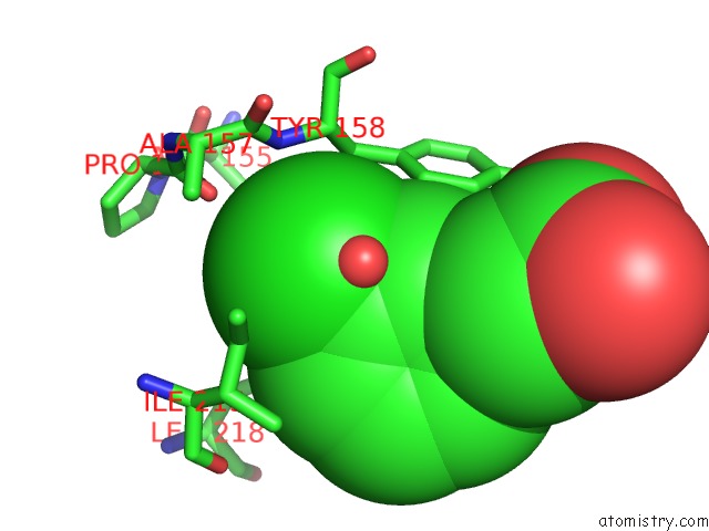
Mono view
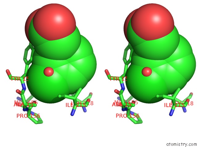
Stereo pair view
Mono view
Stereo pair view
A full contact list of Chlorine with other atoms in the Cl binding
site number 1 of Crystal Structure of M. Tuberculosis Inha in Complex with Nad+ and 3- (3-Chlorophenyl)Propanoic Acid within 5.0Å range:
|
Reference:
M.Sabbah,
V.Mendes,
R.G.Vistal,
D.M.Dias,
M.Zahorszka,
K.Mikusova,
J.Kordulakova,
A.G.Coyne,
T.L.Blundell,
C.Abell.
Fragment-Based Design of Mycobacterium Tuberculosis Inha Inhibitors. J.Med.Chem. 2020.
ISSN: ISSN 0022-2623
PubMed: 32240584
DOI: 10.1021/ACS.JMEDCHEM.0C00007
Page generated: Mon Jul 29 15:07:38 2024
ISSN: ISSN 0022-2623
PubMed: 32240584
DOI: 10.1021/ACS.JMEDCHEM.0C00007
Last articles
Zn in 9J0NZn in 9J0O
Zn in 9J0P
Zn in 9FJX
Zn in 9EKB
Zn in 9C0F
Zn in 9CAH
Zn in 9CH0
Zn in 9CH3
Zn in 9CH1