Chlorine »
PDB 6tma-6ttg »
6tqr »
Chlorine in PDB 6tqr: The Crystal Structure of the Msp Domain of Human Vap-A in Complex with the Phospho-Ffat Motif of STARD3.
Protein crystallography data
The structure of The Crystal Structure of the Msp Domain of Human Vap-A in Complex with the Phospho-Ffat Motif of STARD3., PDB code: 6tqr
was solved by
A.G.Mcewen,
P.Poussin-Courmontagne,
T.Di Mattia,
C.Wendling,
J.Cavarelli,
C.Tomasetto,
F.Alpy,
with X-Ray Crystallography technique. A brief refinement statistics is given in the table below:
| Resolution Low / High (Å) | 42.26 / 1.85 |
| Space group | P 1 |
| Cell size a, b, c (Å), α, β, γ (°) | 39.140, 43.780, 83.640, 89.46, 92.91, 105.13 |
| R / Rfree (%) | 20.4 / 25.4 |
Chlorine Binding Sites:
The binding sites of Chlorine atom in the The Crystal Structure of the Msp Domain of Human Vap-A in Complex with the Phospho-Ffat Motif of STARD3.
(pdb code 6tqr). This binding sites where shown within
5.0 Angstroms radius around Chlorine atom.
In total 7 binding sites of Chlorine where determined in the The Crystal Structure of the Msp Domain of Human Vap-A in Complex with the Phospho-Ffat Motif of STARD3., PDB code: 6tqr:
Jump to Chlorine binding site number: 1; 2; 3; 4; 5; 6; 7;
In total 7 binding sites of Chlorine where determined in the The Crystal Structure of the Msp Domain of Human Vap-A in Complex with the Phospho-Ffat Motif of STARD3., PDB code: 6tqr:
Jump to Chlorine binding site number: 1; 2; 3; 4; 5; 6; 7;
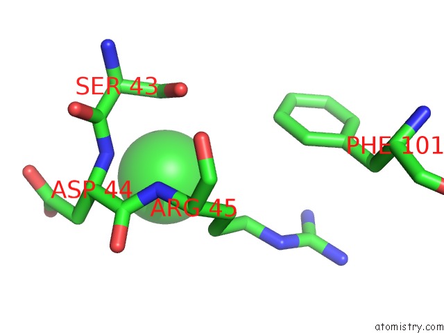
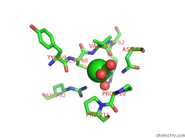
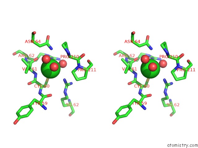
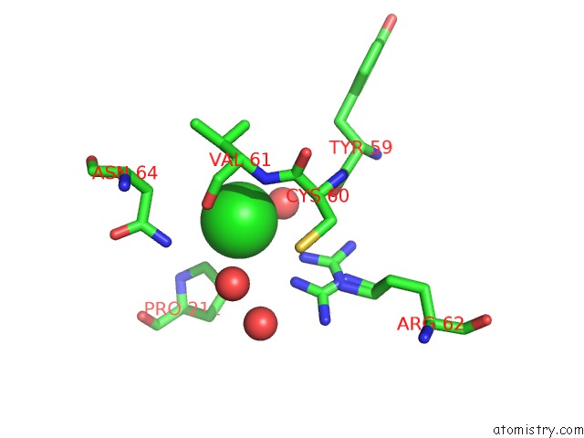
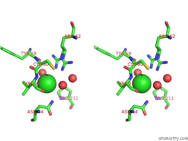
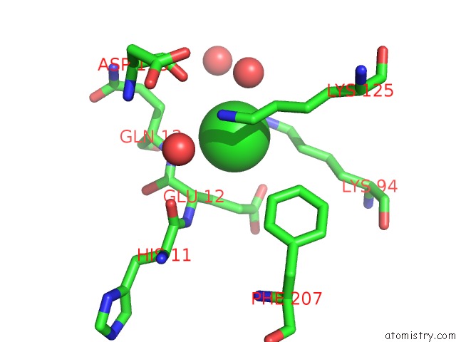
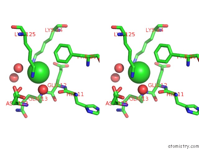
Chlorine binding site 1 out of 7 in 6tqr
Go back to
Chlorine binding site 1 out
of 7 in the The Crystal Structure of the Msp Domain of Human Vap-A in Complex with the Phospho-Ffat Motif of STARD3.
Mono view
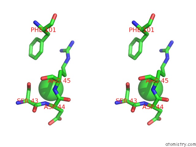
Stereo pair view
Mono view
Stereo pair view
A full contact list of Chlorine with other atoms in the Cl binding
site number 1 of The Crystal Structure of the Msp Domain of Human Vap-A in Complex with the Phospho-Ffat Motif of STARD3. within 5.0Å range:
|
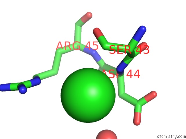
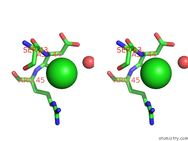
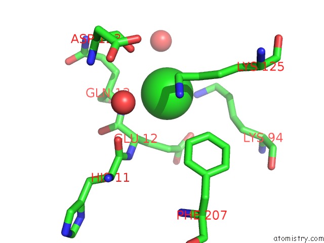
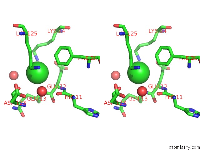
Chlorine binding site 2 out of 7 in 6tqr
Go back to
Chlorine binding site 2 out
of 7 in the The Crystal Structure of the Msp Domain of Human Vap-A in Complex with the Phospho-Ffat Motif of STARD3.
Mono view
Stereo pair view
Mono view
Stereo pair view
A full contact list of Chlorine with other atoms in the Cl binding
site number 2 of The Crystal Structure of the Msp Domain of Human Vap-A in Complex with the Phospho-Ffat Motif of STARD3. within 5.0Å range:
|
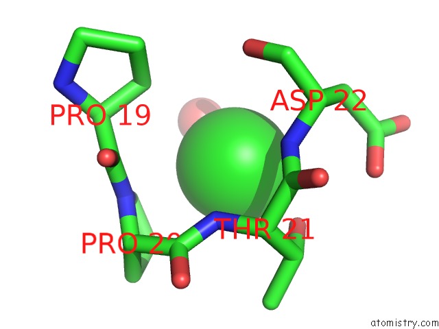
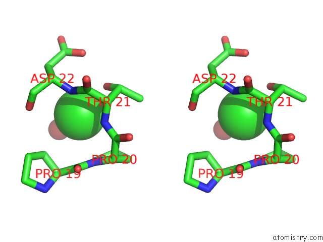
Chlorine binding site 3 out of 7 in 6tqr
Go back to
Chlorine binding site 3 out
of 7 in the The Crystal Structure of the Msp Domain of Human Vap-A in Complex with the Phospho-Ffat Motif of STARD3.
Mono view
Stereo pair view
Mono view
Stereo pair view
A full contact list of Chlorine with other atoms in the Cl binding
site number 3 of The Crystal Structure of the Msp Domain of Human Vap-A in Complex with the Phospho-Ffat Motif of STARD3. within 5.0Å range:
|
Chlorine binding site 4 out of 7 in 6tqr
Go back to
Chlorine binding site 4 out
of 7 in the The Crystal Structure of the Msp Domain of Human Vap-A in Complex with the Phospho-Ffat Motif of STARD3.
Mono view
Stereo pair view
Mono view
Stereo pair view
A full contact list of Chlorine with other atoms in the Cl binding
site number 4 of The Crystal Structure of the Msp Domain of Human Vap-A in Complex with the Phospho-Ffat Motif of STARD3. within 5.0Å range:
|
Chlorine binding site 5 out of 7 in 6tqr
Go back to
Chlorine binding site 5 out
of 7 in the The Crystal Structure of the Msp Domain of Human Vap-A in Complex with the Phospho-Ffat Motif of STARD3.
Mono view
Stereo pair view
Mono view
Stereo pair view
A full contact list of Chlorine with other atoms in the Cl binding
site number 5 of The Crystal Structure of the Msp Domain of Human Vap-A in Complex with the Phospho-Ffat Motif of STARD3. within 5.0Å range:
|
Chlorine binding site 6 out of 7 in 6tqr
Go back to
Chlorine binding site 6 out
of 7 in the The Crystal Structure of the Msp Domain of Human Vap-A in Complex with the Phospho-Ffat Motif of STARD3.
Mono view
Stereo pair view
Mono view
Stereo pair view
A full contact list of Chlorine with other atoms in the Cl binding
site number 6 of The Crystal Structure of the Msp Domain of Human Vap-A in Complex with the Phospho-Ffat Motif of STARD3. within 5.0Å range:
|
Chlorine binding site 7 out of 7 in 6tqr
Go back to
Chlorine binding site 7 out
of 7 in the The Crystal Structure of the Msp Domain of Human Vap-A in Complex with the Phospho-Ffat Motif of STARD3.
Mono view
Stereo pair view
Mono view
Stereo pair view
A full contact list of Chlorine with other atoms in the Cl binding
site number 7 of The Crystal Structure of the Msp Domain of Human Vap-A in Complex with the Phospho-Ffat Motif of STARD3. within 5.0Å range:
|
Reference:
T.Di Mattia,
A.Martinet,
S.Ikhlef,
A.G.Mcewen,
Y.Nomine,
C.Wendling,
P.Poussin-Courmontagne,
L.Voilquin,
P.Eberling,
F.Ruffenach,
J.Cavarelli,
J.Slee,
T.P.Levine,
G.Drin,
C.Tomasetto,
F.Alpy.
Ffat Motif Phosphorylation Controls Formation and Lipid Transfer Function of Inter-Organelle Contacts. Embo J. 04369 2020.
ISSN: ESSN 1460-2075
PubMed: 33124732
DOI: 10.15252/EMBJ.2019104369
Page generated: Mon Jul 29 15:33:50 2024
ISSN: ESSN 1460-2075
PubMed: 33124732
DOI: 10.15252/EMBJ.2019104369
Last articles
Zn in 9J0NZn in 9J0O
Zn in 9J0P
Zn in 9FJX
Zn in 9EKB
Zn in 9C0F
Zn in 9CAH
Zn in 9CH0
Zn in 9CH3
Zn in 9CH1