Chlorine »
PDB 6tun-6u52 »
6tzd »
Chlorine in PDB 6tzd: Crystal Structure of Ketosteroid Isomerase From Pseudomonas Putida (Pksi) Bound to 4-Androstenedione at 280 K
Enzymatic activity of Crystal Structure of Ketosteroid Isomerase From Pseudomonas Putida (Pksi) Bound to 4-Androstenedione at 280 K
All present enzymatic activity of Crystal Structure of Ketosteroid Isomerase From Pseudomonas Putida (Pksi) Bound to 4-Androstenedione at 280 K:
5.3.3.1;
5.3.3.1;
Protein crystallography data
The structure of Crystal Structure of Ketosteroid Isomerase From Pseudomonas Putida (Pksi) Bound to 4-Androstenedione at 280 K, PDB code: 6tzd
was solved by
F.Yabukarski,
D.Herschlag,
with X-Ray Crystallography technique. A brief refinement statistics is given in the table below:
| Resolution Low / High (Å) | 34.66 / 1.45 |
| Space group | P 21 21 21 |
| Cell size a, b, c (Å), α, β, γ (°) | 36.229, 74.385, 95.345, 90.00, 90.00, 90.00 |
| R / Rfree (%) | 14.6 / 17.4 |
Other elements in 6tzd:
The structure of Crystal Structure of Ketosteroid Isomerase From Pseudomonas Putida (Pksi) Bound to 4-Androstenedione at 280 K also contains other interesting chemical elements:
| Magnesium | (Mg) | 1 atom |
Chlorine Binding Sites:
The binding sites of Chlorine atom in the Crystal Structure of Ketosteroid Isomerase From Pseudomonas Putida (Pksi) Bound to 4-Androstenedione at 280 K
(pdb code 6tzd). This binding sites where shown within
5.0 Angstroms radius around Chlorine atom.
In total 2 binding sites of Chlorine where determined in the Crystal Structure of Ketosteroid Isomerase From Pseudomonas Putida (Pksi) Bound to 4-Androstenedione at 280 K, PDB code: 6tzd:
Jump to Chlorine binding site number: 1; 2;
In total 2 binding sites of Chlorine where determined in the Crystal Structure of Ketosteroid Isomerase From Pseudomonas Putida (Pksi) Bound to 4-Androstenedione at 280 K, PDB code: 6tzd:
Jump to Chlorine binding site number: 1; 2;
Chlorine binding site 1 out of 2 in 6tzd
Go back to
Chlorine binding site 1 out
of 2 in the Crystal Structure of Ketosteroid Isomerase From Pseudomonas Putida (Pksi) Bound to 4-Androstenedione at 280 K
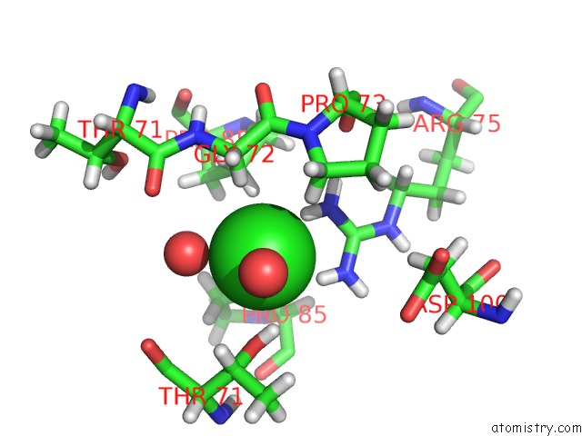
Mono view
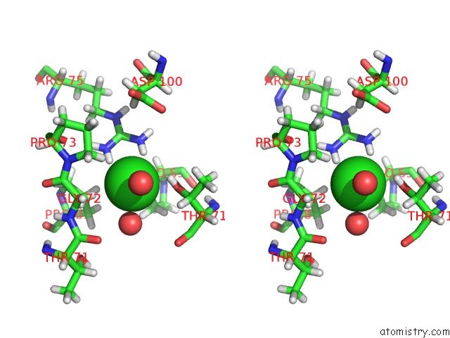
Stereo pair view
Mono view
Stereo pair view
A full contact list of Chlorine with other atoms in the Cl binding
site number 1 of Crystal Structure of Ketosteroid Isomerase From Pseudomonas Putida (Pksi) Bound to 4-Androstenedione at 280 K within 5.0Å range:
|
Chlorine binding site 2 out of 2 in 6tzd
Go back to
Chlorine binding site 2 out
of 2 in the Crystal Structure of Ketosteroid Isomerase From Pseudomonas Putida (Pksi) Bound to 4-Androstenedione at 280 K
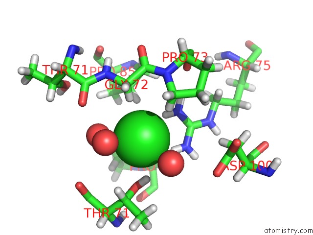
Mono view
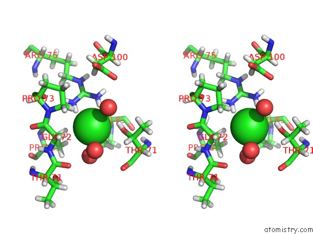
Stereo pair view
Mono view
Stereo pair view
A full contact list of Chlorine with other atoms in the Cl binding
site number 2 of Crystal Structure of Ketosteroid Isomerase From Pseudomonas Putida (Pksi) Bound to 4-Androstenedione at 280 K within 5.0Å range:
|
Reference:
F.Yabukarski,
D.Herschlag.
Assessing Active Site Positioning and Testing Catalytic Proposals Via Ketosteroid Isomerase Conformational Ensembles To Be Published.
Page generated: Sat Jul 12 20:20:30 2025
Last articles
F in 7QV9F in 7QSC
F in 7QTM
F in 7QTV
F in 7QU0
F in 7QRA
F in 7QRB
F in 7QRC
F in 7QR9
F in 7QQ6