Chlorine »
PDB 6tun-6u52 »
6u2m »
Chlorine in PDB 6u2m: Crystal Structure of A Halotag-Based Calcium Indicator, Halocamp V2, Bound to JF635
Protein crystallography data
The structure of Crystal Structure of A Halotag-Based Calcium Indicator, Halocamp V2, Bound to JF635, PDB code: 6u2m
was solved by
C.Deo,
E.R.Schreiter,
with X-Ray Crystallography technique. A brief refinement statistics is given in the table below:
| Resolution Low / High (Å) | 122.58 / 2.00 |
| Space group | P 1 2 1 |
| Cell size a, b, c (Å), α, β, γ (°) | 92.560, 60.662, 122.600, 90.00, 91.02, 90.00 |
| R / Rfree (%) | 18.6 / 22.6 |
Other elements in 6u2m:
The structure of Crystal Structure of A Halotag-Based Calcium Indicator, Halocamp V2, Bound to JF635 also contains other interesting chemical elements:
| Fluorine | (F) | 4 atoms |
| Silicon | (Si) | 2 atoms |
| Calcium | (Ca) | 8 atoms |
Chlorine Binding Sites:
The binding sites of Chlorine atom in the Crystal Structure of A Halotag-Based Calcium Indicator, Halocamp V2, Bound to JF635
(pdb code 6u2m). This binding sites where shown within
5.0 Angstroms radius around Chlorine atom.
In total 2 binding sites of Chlorine where determined in the Crystal Structure of A Halotag-Based Calcium Indicator, Halocamp V2, Bound to JF635, PDB code: 6u2m:
Jump to Chlorine binding site number: 1; 2;
In total 2 binding sites of Chlorine where determined in the Crystal Structure of A Halotag-Based Calcium Indicator, Halocamp V2, Bound to JF635, PDB code: 6u2m:
Jump to Chlorine binding site number: 1; 2;
Chlorine binding site 1 out of 2 in 6u2m
Go back to
Chlorine binding site 1 out
of 2 in the Crystal Structure of A Halotag-Based Calcium Indicator, Halocamp V2, Bound to JF635
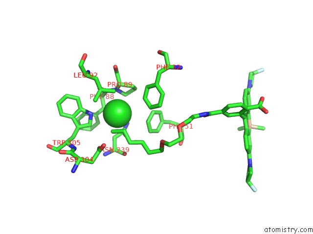
Mono view
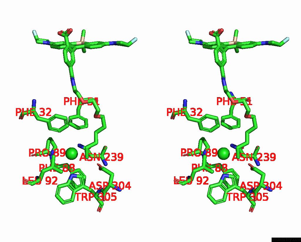
Stereo pair view
Mono view
Stereo pair view
A full contact list of Chlorine with other atoms in the Cl binding
site number 1 of Crystal Structure of A Halotag-Based Calcium Indicator, Halocamp V2, Bound to JF635 within 5.0Å range:
|
Chlorine binding site 2 out of 2 in 6u2m
Go back to
Chlorine binding site 2 out
of 2 in the Crystal Structure of A Halotag-Based Calcium Indicator, Halocamp V2, Bound to JF635
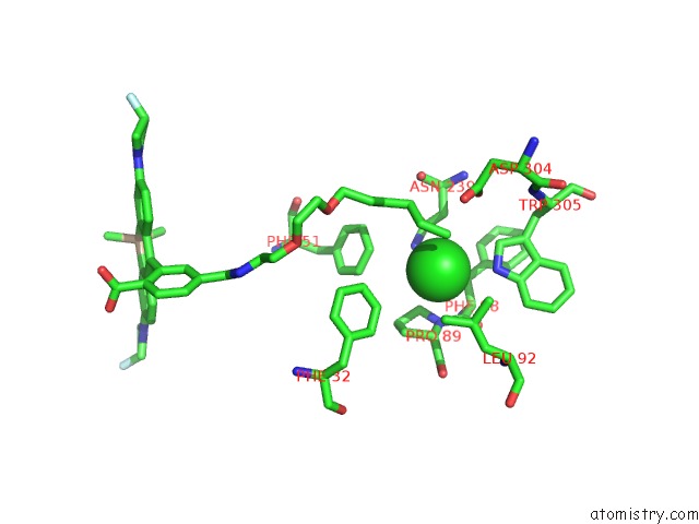
Mono view
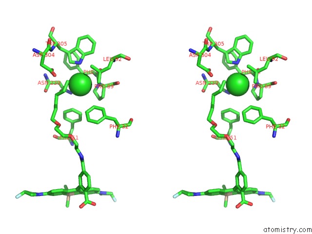
Stereo pair view
Mono view
Stereo pair view
A full contact list of Chlorine with other atoms in the Cl binding
site number 2 of Crystal Structure of A Halotag-Based Calcium Indicator, Halocamp V2, Bound to JF635 within 5.0Å range:
|
Reference:
C.Deo,
L.D.Lavis,
E.R.Schreiter.
Crystal Structure of A Halotag-Based Calcium Indicator, Halocamp V2 To Be Published.
Page generated: Sat Jul 12 20:22:42 2025
Last articles
F in 4L6QF in 4L7F
F in 4L4L
F in 4L46
F in 4L45
F in 4L44
F in 4L43
F in 4L42
F in 4L3L
F in 4L3J