Chlorine »
PDB 6u5y-6udu »
6ub6 »
Chlorine in PDB 6ub6: Crystal Structure of A GH128 (Subgroup IV) Endo-Beta-1,3-Glucanase From Lentinula Edodes (LEGH128_IV) in Complex with Laminaritetraose
Protein crystallography data
The structure of Crystal Structure of A GH128 (Subgroup IV) Endo-Beta-1,3-Glucanase From Lentinula Edodes (LEGH128_IV) in Complex with Laminaritetraose, PDB code: 6ub6
was solved by
C.R.Santos,
E.A.Lima,
F.Mandelli,
M.T.Murakami,
with X-Ray Crystallography technique. A brief refinement statistics is given in the table below:
| Resolution Low / High (Å) | 49.25 / 1.25 |
| Space group | P 1 21 1 |
| Cell size a, b, c (Å), α, β, γ (°) | 45.871, 47.678, 52.549, 90.00, 110.42, 90.00 |
| R / Rfree (%) | 17.4 / 19.1 |
Chlorine Binding Sites:
The binding sites of Chlorine atom in the Crystal Structure of A GH128 (Subgroup IV) Endo-Beta-1,3-Glucanase From Lentinula Edodes (LEGH128_IV) in Complex with Laminaritetraose
(pdb code 6ub6). This binding sites where shown within
5.0 Angstroms radius around Chlorine atom.
In total only one binding site of Chlorine was determined in the Crystal Structure of A GH128 (Subgroup IV) Endo-Beta-1,3-Glucanase From Lentinula Edodes (LEGH128_IV) in Complex with Laminaritetraose, PDB code: 6ub6:
In total only one binding site of Chlorine was determined in the Crystal Structure of A GH128 (Subgroup IV) Endo-Beta-1,3-Glucanase From Lentinula Edodes (LEGH128_IV) in Complex with Laminaritetraose, PDB code: 6ub6:
Chlorine binding site 1 out of 1 in 6ub6
Go back to
Chlorine binding site 1 out
of 1 in the Crystal Structure of A GH128 (Subgroup IV) Endo-Beta-1,3-Glucanase From Lentinula Edodes (LEGH128_IV) in Complex with Laminaritetraose
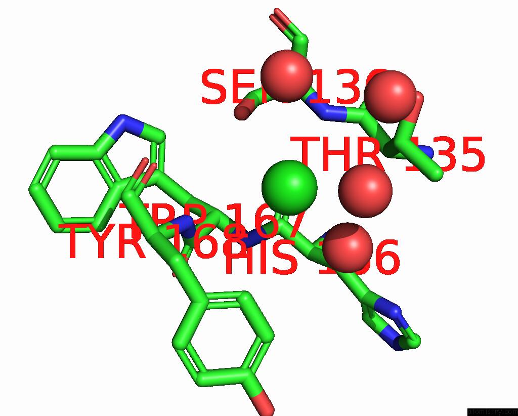
Mono view
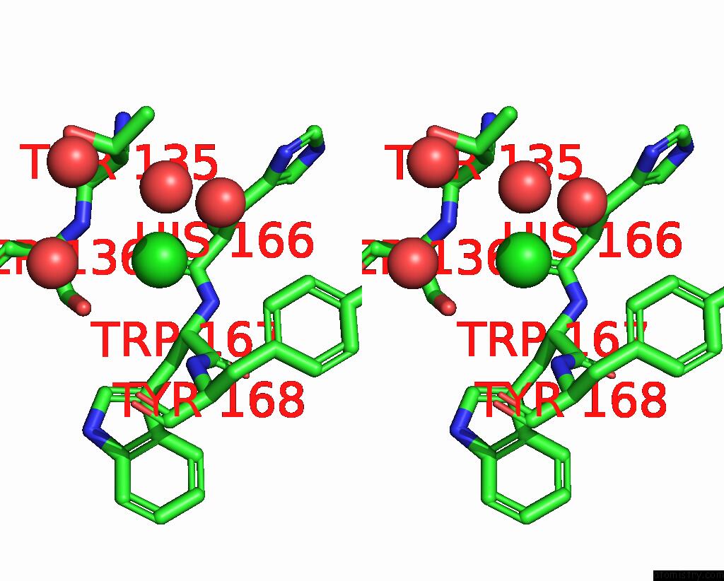
Stereo pair view
Mono view
Stereo pair view
A full contact list of Chlorine with other atoms in the Cl binding
site number 1 of Crystal Structure of A GH128 (Subgroup IV) Endo-Beta-1,3-Glucanase From Lentinula Edodes (LEGH128_IV) in Complex with Laminaritetraose within 5.0Å range:
|
Reference:
C.R.Santos,
E.A.Lima,
F.Mandelli,
M.T.Murakami.
Structural Insights Into Beta-1,3-Glucan Cleavage By A Glycoside Hydrolase Family Nat.Chem.Biol. 2020.
ISSN: ESSN 1552-4469
DOI: 10.1038/S41589-020-0554-5
Page generated: Sat Jul 12 20:28:23 2025
ISSN: ESSN 1552-4469
DOI: 10.1038/S41589-020-0554-5
Last articles
Cl in 8C1HCl in 8C14
Cl in 8C1I
Cl in 8C1G
Cl in 8C1F
Cl in 8C1E
Cl in 8C1D
Cl in 8C15
Cl in 8C04
Cl in 8C0K