Chlorine »
PDB 6u5y-6udu »
6ubq »
Chlorine in PDB 6ubq: Crystal Structure of Ketosteroid Isomerase From Pseudomonas Putida (Pksi) Bound to 4-Androstenedione at 100 K
Enzymatic activity of Crystal Structure of Ketosteroid Isomerase From Pseudomonas Putida (Pksi) Bound to 4-Androstenedione at 100 K
All present enzymatic activity of Crystal Structure of Ketosteroid Isomerase From Pseudomonas Putida (Pksi) Bound to 4-Androstenedione at 100 K:
5.3.3.1;
5.3.3.1;
Protein crystallography data
The structure of Crystal Structure of Ketosteroid Isomerase From Pseudomonas Putida (Pksi) Bound to 4-Androstenedione at 100 K, PDB code: 6ubq
was solved by
F.Yabukarski,
D.Herschlag,
with X-Ray Crystallography technique. A brief refinement statistics is given in the table below:
| Resolution Low / High (Å) | 34.64 / 1.30 |
| Space group | P 21 21 21 |
| Cell size a, b, c (Å), α, β, γ (°) | 36.209, 74.316, 95.562, 90.00, 90.00, 90.00 |
| R / Rfree (%) | 15.2 / 17 |
Other elements in 6ubq:
The structure of Crystal Structure of Ketosteroid Isomerase From Pseudomonas Putida (Pksi) Bound to 4-Androstenedione at 100 K also contains other interesting chemical elements:
| Magnesium | (Mg) | 1 atom |
Chlorine Binding Sites:
The binding sites of Chlorine atom in the Crystal Structure of Ketosteroid Isomerase From Pseudomonas Putida (Pksi) Bound to 4-Androstenedione at 100 K
(pdb code 6ubq). This binding sites where shown within
5.0 Angstroms radius around Chlorine atom.
In total 2 binding sites of Chlorine where determined in the Crystal Structure of Ketosteroid Isomerase From Pseudomonas Putida (Pksi) Bound to 4-Androstenedione at 100 K, PDB code: 6ubq:
Jump to Chlorine binding site number: 1; 2;
In total 2 binding sites of Chlorine where determined in the Crystal Structure of Ketosteroid Isomerase From Pseudomonas Putida (Pksi) Bound to 4-Androstenedione at 100 K, PDB code: 6ubq:
Jump to Chlorine binding site number: 1; 2;
Chlorine binding site 1 out of 2 in 6ubq
Go back to
Chlorine binding site 1 out
of 2 in the Crystal Structure of Ketosteroid Isomerase From Pseudomonas Putida (Pksi) Bound to 4-Androstenedione at 100 K
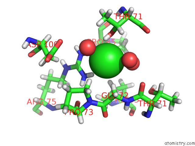
Mono view
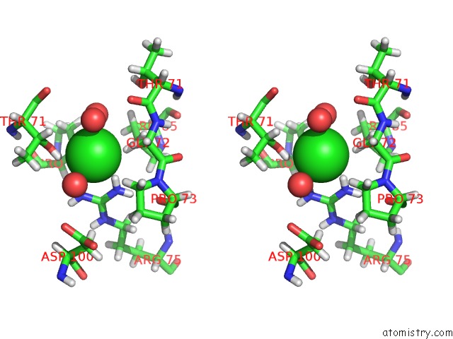
Stereo pair view
Mono view
Stereo pair view
A full contact list of Chlorine with other atoms in the Cl binding
site number 1 of Crystal Structure of Ketosteroid Isomerase From Pseudomonas Putida (Pksi) Bound to 4-Androstenedione at 100 K within 5.0Å range:
|
Chlorine binding site 2 out of 2 in 6ubq
Go back to
Chlorine binding site 2 out
of 2 in the Crystal Structure of Ketosteroid Isomerase From Pseudomonas Putida (Pksi) Bound to 4-Androstenedione at 100 K
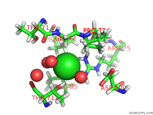
Mono view
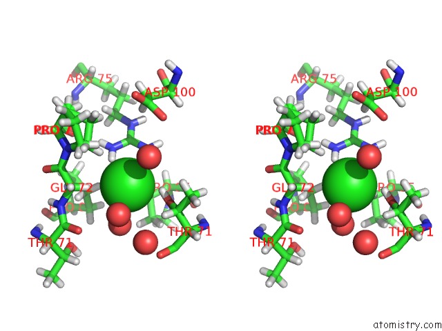
Stereo pair view
Mono view
Stereo pair view
A full contact list of Chlorine with other atoms in the Cl binding
site number 2 of Crystal Structure of Ketosteroid Isomerase From Pseudomonas Putida (Pksi) Bound to 4-Androstenedione at 100 K within 5.0Å range:
|
Reference:
F.Yabukarski,
D.Herschlag.
Assessing Active Site Positioning and Testing Catalytic Proposals Via Ketosteroid Isomerase Conformational Ensembles To Be Published.
Page generated: Sat Jul 12 20:28:31 2025
Last articles
Fe in 2YXOFe in 2YRS
Fe in 2YXC
Fe in 2YNM
Fe in 2YVJ
Fe in 2YP1
Fe in 2YU2
Fe in 2YU1
Fe in 2YQB
Fe in 2YOO