Chlorine »
PDB 6vck-6vkd »
6vcl »
Chlorine in PDB 6vcl: Crystal Structure of E.Coli Rpph-Dapf in Complex with Pppgpp, MG2+ and F-
Enzymatic activity of Crystal Structure of E.Coli Rpph-Dapf in Complex with Pppgpp, MG2+ and F-
All present enzymatic activity of Crystal Structure of E.Coli Rpph-Dapf in Complex with Pppgpp, MG2+ and F-:
5.1.1.7;
5.1.1.7;
Protein crystallography data
The structure of Crystal Structure of E.Coli Rpph-Dapf in Complex with Pppgpp, MG2+ and F-, PDB code: 6vcl
was solved by
A.Gao,
N.Vasilyev,
A.Kaushik,
W.Duan,
A.Serganov,
with X-Ray Crystallography technique. A brief refinement statistics is given in the table below:
| Resolution Low / High (Å) | 30.00 / 2.06 |
| Space group | C 2 2 21 |
| Cell size a, b, c (Å), α, β, γ (°) | 162.444, 190.649, 51.336, 90.00, 90.00, 90.00 |
| R / Rfree (%) | 19.1 / 22.7 |
Other elements in 6vcl:
The structure of Crystal Structure of E.Coli Rpph-Dapf in Complex with Pppgpp, MG2+ and F- also contains other interesting chemical elements:
| Fluorine | (F) | 1 atom |
| Magnesium | (Mg) | 4 atoms |
Chlorine Binding Sites:
The binding sites of Chlorine atom in the Crystal Structure of E.Coli Rpph-Dapf in Complex with Pppgpp, MG2+ and F-
(pdb code 6vcl). This binding sites where shown within
5.0 Angstroms radius around Chlorine atom.
In total 2 binding sites of Chlorine where determined in the Crystal Structure of E.Coli Rpph-Dapf in Complex with Pppgpp, MG2+ and F-, PDB code: 6vcl:
Jump to Chlorine binding site number: 1; 2;
In total 2 binding sites of Chlorine where determined in the Crystal Structure of E.Coli Rpph-Dapf in Complex with Pppgpp, MG2+ and F-, PDB code: 6vcl:
Jump to Chlorine binding site number: 1; 2;
Chlorine binding site 1 out of 2 in 6vcl
Go back to
Chlorine binding site 1 out
of 2 in the Crystal Structure of E.Coli Rpph-Dapf in Complex with Pppgpp, MG2+ and F-
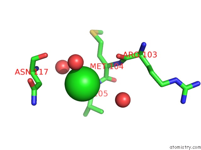
Mono view
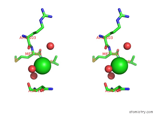
Stereo pair view
Mono view
Stereo pair view
A full contact list of Chlorine with other atoms in the Cl binding
site number 1 of Crystal Structure of E.Coli Rpph-Dapf in Complex with Pppgpp, MG2+ and F- within 5.0Å range:
|
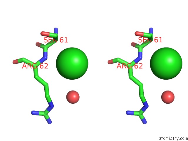
Chlorine binding site 2 out of 2 in 6vcl
Go back to
Chlorine binding site 2 out
of 2 in the Crystal Structure of E.Coli Rpph-Dapf in Complex with Pppgpp, MG2+ and F-
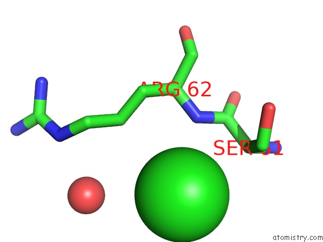
Mono view
Stereo pair view
Mono view
Stereo pair view
A full contact list of Chlorine with other atoms in the Cl binding
site number 2 of Crystal Structure of E.Coli Rpph-Dapf in Complex with Pppgpp, MG2+ and F- within 5.0Å range:
|
Reference:
A.Gao,
N.Vasilyev,
A.Kaushik,
W.Duan,
A.Serganov.
Principles of Rna and Nucleotide Discrimination By the Rna Processing Enzyme Rpph. Nucleic Acids Res. 2020.
ISSN: ESSN 1362-4962
PubMed: 31960065
DOI: 10.1093/NAR/GKAA024
Page generated: Mon Jul 29 16:14:58 2024
ISSN: ESSN 1362-4962
PubMed: 31960065
DOI: 10.1093/NAR/GKAA024
Last articles
Zn in 9J0NZn in 9J0O
Zn in 9J0P
Zn in 9FJX
Zn in 9EKB
Zn in 9C0F
Zn in 9CAH
Zn in 9CH0
Zn in 9CH3
Zn in 9CH1