Chlorine »
PDB 6vck-6vkd »
6vcw »
Chlorine in PDB 6vcw: Crystal Structure of Medicago Truncatula S-Adenosylmethionine Synthase 3A (MTMAT3A)
Enzymatic activity of Crystal Structure of Medicago Truncatula S-Adenosylmethionine Synthase 3A (MTMAT3A)
All present enzymatic activity of Crystal Structure of Medicago Truncatula S-Adenosylmethionine Synthase 3A (MTMAT3A):
2.5.1.6;
2.5.1.6;
Protein crystallography data
The structure of Crystal Structure of Medicago Truncatula S-Adenosylmethionine Synthase 3A (MTMAT3A), PDB code: 6vcw
was solved by
B.Sekula,
M.Ruszkowski,
Z.Dauter,
with X-Ray Crystallography technique. A brief refinement statistics is given in the table below:
| Resolution Low / High (Å) | 48.99 / 1.40 |
| Space group | C 1 2 1 |
| Cell size a, b, c (Å), α, β, γ (°) | 111.602, 62.042, 102.284, 90.00, 90.02, 90.00 |
| R / Rfree (%) | 11.5 / 15.9 |
Other elements in 6vcw:
The structure of Crystal Structure of Medicago Truncatula S-Adenosylmethionine Synthase 3A (MTMAT3A) also contains other interesting chemical elements:
| Magnesium | (Mg) | 4 atoms |
Chlorine Binding Sites:
The binding sites of Chlorine atom in the Crystal Structure of Medicago Truncatula S-Adenosylmethionine Synthase 3A (MTMAT3A)
(pdb code 6vcw). This binding sites where shown within
5.0 Angstroms radius around Chlorine atom.
In total 2 binding sites of Chlorine where determined in the Crystal Structure of Medicago Truncatula S-Adenosylmethionine Synthase 3A (MTMAT3A), PDB code: 6vcw:
Jump to Chlorine binding site number: 1; 2;
In total 2 binding sites of Chlorine where determined in the Crystal Structure of Medicago Truncatula S-Adenosylmethionine Synthase 3A (MTMAT3A), PDB code: 6vcw:
Jump to Chlorine binding site number: 1; 2;
Chlorine binding site 1 out of 2 in 6vcw
Go back to
Chlorine binding site 1 out
of 2 in the Crystal Structure of Medicago Truncatula S-Adenosylmethionine Synthase 3A (MTMAT3A)
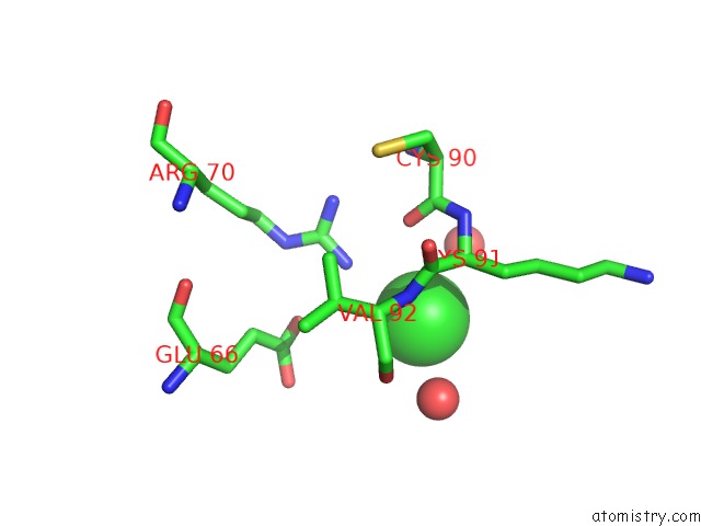
Mono view
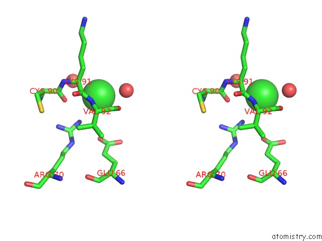
Stereo pair view
Mono view
Stereo pair view
A full contact list of Chlorine with other atoms in the Cl binding
site number 1 of Crystal Structure of Medicago Truncatula S-Adenosylmethionine Synthase 3A (MTMAT3A) within 5.0Å range:
|
Chlorine binding site 2 out of 2 in 6vcw
Go back to
Chlorine binding site 2 out
of 2 in the Crystal Structure of Medicago Truncatula S-Adenosylmethionine Synthase 3A (MTMAT3A)
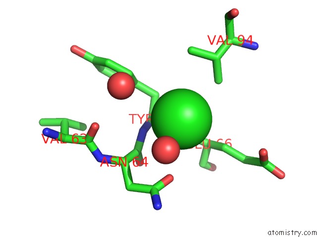
Mono view
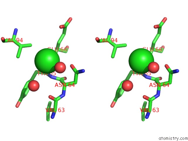
Stereo pair view
Mono view
Stereo pair view
A full contact list of Chlorine with other atoms in the Cl binding
site number 2 of Crystal Structure of Medicago Truncatula S-Adenosylmethionine Synthase 3A (MTMAT3A) within 5.0Å range:
|
Reference:
B.Sekula,
M.Ruszkowski,
Z.Dauter.
S-Adenosylmethionine Synthases in Plants: Structural Characterization of Type I and II Isoenzymes From Arabidopsis Thaliana and Medicago Truncatula. Int.J.Biol.Macromol. 2020.
ISSN: ISSN 0141-8130
PubMed: 32057875
DOI: 10.1016/J.IJBIOMAC.2020.02.100
Page generated: Mon Jul 29 16:14:58 2024
ISSN: ISSN 0141-8130
PubMed: 32057875
DOI: 10.1016/J.IJBIOMAC.2020.02.100
Last articles
Zn in 9J0NZn in 9J0O
Zn in 9J0P
Zn in 9FJX
Zn in 9EKB
Zn in 9C0F
Zn in 9CAH
Zn in 9CH0
Zn in 9CH3
Zn in 9CH1