Chlorine »
PDB 6vke-6vww »
6vlt »
Chlorine in PDB 6vlt: Crystal Structure of Human P450 2C9*2 Genetic Variant in Complex with Losartan
Enzymatic activity of Crystal Structure of Human P450 2C9*2 Genetic Variant in Complex with Losartan
All present enzymatic activity of Crystal Structure of Human P450 2C9*2 Genetic Variant in Complex with Losartan:
1.14.14.1; 1.14.14.51; 1.14.14.52; 1.14.14.53;
1.14.14.1; 1.14.14.51; 1.14.14.52; 1.14.14.53;
Protein crystallography data
The structure of Crystal Structure of Human P450 2C9*2 Genetic Variant in Complex with Losartan, PDB code: 6vlt
was solved by
M.B.Shah,
with X-Ray Crystallography technique. A brief refinement statistics is given in the table below:
| Resolution Low / High (Å) | 50.01 / 3.12 |
| Space group | P 31 |
| Cell size a, b, c (Å), α, β, γ (°) | 238.100, 238.100, 109.851, 90.00, 90.00, 120.00 |
| R / Rfree (%) | 15.6 / 22.1 |
Other elements in 6vlt:
The structure of Crystal Structure of Human P450 2C9*2 Genetic Variant in Complex with Losartan also contains other interesting chemical elements:
| Iron | (Fe) | 8 atoms |
Chlorine Binding Sites:
The binding sites of Chlorine atom in the Crystal Structure of Human P450 2C9*2 Genetic Variant in Complex with Losartan
(pdb code 6vlt). This binding sites where shown within
5.0 Angstroms radius around Chlorine atom.
In total 8 binding sites of Chlorine where determined in the Crystal Structure of Human P450 2C9*2 Genetic Variant in Complex with Losartan, PDB code: 6vlt:
Jump to Chlorine binding site number: 1; 2; 3; 4; 5; 6; 7; 8;
In total 8 binding sites of Chlorine where determined in the Crystal Structure of Human P450 2C9*2 Genetic Variant in Complex with Losartan, PDB code: 6vlt:
Jump to Chlorine binding site number: 1; 2; 3; 4; 5; 6; 7; 8;
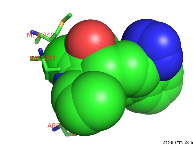
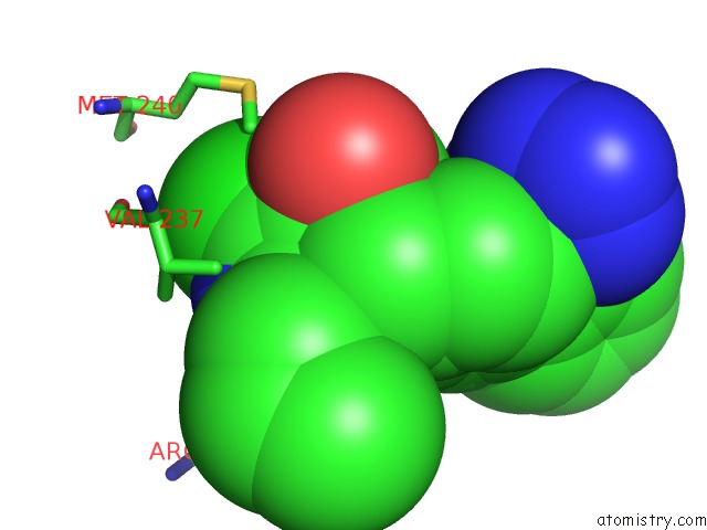
Chlorine binding site 1 out of 8 in 6vlt
Go back to
Chlorine binding site 1 out
of 8 in the Crystal Structure of Human P450 2C9*2 Genetic Variant in Complex with Losartan

Mono view

Stereo pair view
Mono view
Stereo pair view
A full contact list of Chlorine with other atoms in the Cl binding
site number 1 of Crystal Structure of Human P450 2C9*2 Genetic Variant in Complex with Losartan within 5.0Å range:
|
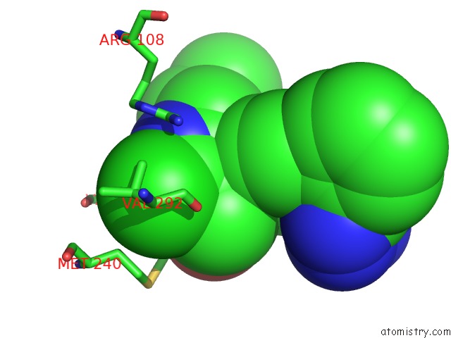
Chlorine binding site 2 out of 8 in 6vlt
Go back to
Chlorine binding site 2 out
of 8 in the Crystal Structure of Human P450 2C9*2 Genetic Variant in Complex with Losartan

Mono view

Stereo pair view
Mono view
Stereo pair view
A full contact list of Chlorine with other atoms in the Cl binding
site number 2 of Crystal Structure of Human P450 2C9*2 Genetic Variant in Complex with Losartan within 5.0Å range:
|
Chlorine binding site 3 out of 8 in 6vlt
Go back to
Chlorine binding site 3 out
of 8 in the Crystal Structure of Human P450 2C9*2 Genetic Variant in Complex with Losartan

Mono view

Stereo pair view
Mono view
Stereo pair view
A full contact list of Chlorine with other atoms in the Cl binding
site number 3 of Crystal Structure of Human P450 2C9*2 Genetic Variant in Complex with Losartan within 5.0Å range:
|
Chlorine binding site 4 out of 8 in 6vlt
Go back to
Chlorine binding site 4 out
of 8 in the Crystal Structure of Human P450 2C9*2 Genetic Variant in Complex with Losartan

Mono view

Stereo pair view
Mono view
Stereo pair view
A full contact list of Chlorine with other atoms in the Cl binding
site number 4 of Crystal Structure of Human P450 2C9*2 Genetic Variant in Complex with Losartan within 5.0Å range:
|
Chlorine binding site 5 out of 8 in 6vlt
Go back to
Chlorine binding site 5 out
of 8 in the Crystal Structure of Human P450 2C9*2 Genetic Variant in Complex with Losartan
Mono view

Stereo pair view
Mono view
Stereo pair view
A full contact list of Chlorine with other atoms in the Cl binding
site number 5 of Crystal Structure of Human P450 2C9*2 Genetic Variant in Complex with Losartan within 5.0Å range:
|
Chlorine binding site 6 out of 8 in 6vlt
Go back to
Chlorine binding site 6 out
of 8 in the Crystal Structure of Human P450 2C9*2 Genetic Variant in Complex with Losartan

Mono view

Stereo pair view
Mono view
Stereo pair view
A full contact list of Chlorine with other atoms in the Cl binding
site number 6 of Crystal Structure of Human P450 2C9*2 Genetic Variant in Complex with Losartan within 5.0Å range:
|
Chlorine binding site 7 out of 8 in 6vlt
Go back to
Chlorine binding site 7 out
of 8 in the Crystal Structure of Human P450 2C9*2 Genetic Variant in Complex with Losartan
Mono view

Stereo pair view
Mono view
Stereo pair view
A full contact list of Chlorine with other atoms in the Cl binding
site number 7 of Crystal Structure of Human P450 2C9*2 Genetic Variant in Complex with Losartan within 5.0Å range:
|
Chlorine binding site 8 out of 8 in 6vlt
Go back to
Chlorine binding site 8 out
of 8 in the Crystal Structure of Human P450 2C9*2 Genetic Variant in Complex with Losartan
Mono view

Stereo pair view
Mono view
Stereo pair view
A full contact list of Chlorine with other atoms in the Cl binding
site number 8 of Crystal Structure of Human P450 2C9*2 Genetic Variant in Complex with Losartan within 5.0Å range:
|
Reference:
S.J.Parikh,
C.M.Evans,
J.O.Obi,
Q.Zhang,
K.Maekawa,
K.C.Glass,
M.B.Shah.
Structure of Cytochrome P450 2C9*2 in Complex with Losartan: Insights Into the Effect of Genetic Polymorphism. Mol.Pharmacol. V. 98 529 2020.
ISSN: ESSN 1521-0111
PubMed: 32938720
DOI: 10.1124/MOLPHARM.120.000042
Page generated: Mon Jul 29 16:24:23 2024
ISSN: ESSN 1521-0111
PubMed: 32938720
DOI: 10.1124/MOLPHARM.120.000042
Last articles
Zn in 9J0NZn in 9J0O
Zn in 9J0P
Zn in 9FJX
Zn in 9EKB
Zn in 9C0F
Zn in 9CAH
Zn in 9CH0
Zn in 9CH3
Zn in 9CH1