Chlorine »
PDB 6w7p-6wji »
6wie »
Chlorine in PDB 6wie: Post-Catalytic Nicked Complex of Human Polymerase Mu on A Complementary Dna Double-Strand Break Substrate
Enzymatic activity of Post-Catalytic Nicked Complex of Human Polymerase Mu on A Complementary Dna Double-Strand Break Substrate
All present enzymatic activity of Post-Catalytic Nicked Complex of Human Polymerase Mu on A Complementary Dna Double-Strand Break Substrate:
2.7.7.7;
2.7.7.7;
Protein crystallography data
The structure of Post-Catalytic Nicked Complex of Human Polymerase Mu on A Complementary Dna Double-Strand Break Substrate, PDB code: 6wie
was solved by
A.M.Kaminski,
T.A.Kunkel,
L.C.Pedersen,
K.Bebenek,
with X-Ray Crystallography technique. A brief refinement statistics is given in the table below:
| Resolution Low / High (Å) | 34.90 / 1.50 |
| Space group | P 21 21 21 |
| Cell size a, b, c (Å), α, β, γ (°) | 60.073, 62.247, 118.318, 90.00, 90.00, 90.00 |
| R / Rfree (%) | 17 / 18.7 |
Other elements in 6wie:
The structure of Post-Catalytic Nicked Complex of Human Polymerase Mu on A Complementary Dna Double-Strand Break Substrate also contains other interesting chemical elements:
| Magnesium | (Mg) | 2 atoms |
| Sodium | (Na) | 2 atoms |
Chlorine Binding Sites:
The binding sites of Chlorine atom in the Post-Catalytic Nicked Complex of Human Polymerase Mu on A Complementary Dna Double-Strand Break Substrate
(pdb code 6wie). This binding sites where shown within
5.0 Angstroms radius around Chlorine atom.
In total only one binding site of Chlorine was determined in the Post-Catalytic Nicked Complex of Human Polymerase Mu on A Complementary Dna Double-Strand Break Substrate, PDB code: 6wie:
In total only one binding site of Chlorine was determined in the Post-Catalytic Nicked Complex of Human Polymerase Mu on A Complementary Dna Double-Strand Break Substrate, PDB code: 6wie:
Chlorine binding site 1 out of 1 in 6wie
Go back to
Chlorine binding site 1 out
of 1 in the Post-Catalytic Nicked Complex of Human Polymerase Mu on A Complementary Dna Double-Strand Break Substrate
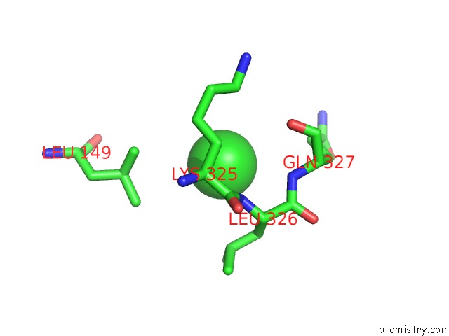
Mono view
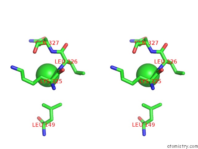
Stereo pair view
Mono view
Stereo pair view
A full contact list of Chlorine with other atoms in the Cl binding
site number 1 of Post-Catalytic Nicked Complex of Human Polymerase Mu on A Complementary Dna Double-Strand Break Substrate within 5.0Å range:
|
Reference:
A.M.Kaminski,
J.M.Pryor,
D.A.Ramsden,
T.A.Kunkel,
L.C.Pedersen,
K.Bebenek.
Structural Snapshots of Human Dna Polymerase Mu Engaged on A Dna Double-Strand Break. Nat Commun V. 11 4784 2020.
ISSN: ESSN 2041-1723
PubMed: 32963245
DOI: 10.1038/S41467-020-18506-5
Page generated: Mon Jul 29 16:45:48 2024
ISSN: ESSN 2041-1723
PubMed: 32963245
DOI: 10.1038/S41467-020-18506-5
Last articles
Zn in 9J0NZn in 9J0O
Zn in 9J0P
Zn in 9FJX
Zn in 9EKB
Zn in 9C0F
Zn in 9CAH
Zn in 9CH0
Zn in 9CH3
Zn in 9CH1