Chlorine »
PDB 6wko-6wv7 »
6wma »
Chlorine in PDB 6wma: Crystal Structure of A Soluble Variant of Full-Length Human APOBEC3G (pH 7.6)
Protein crystallography data
The structure of Crystal Structure of A Soluble Variant of Full-Length Human APOBEC3G (pH 7.6), PDB code: 6wma
was solved by
A.Maiti,
H.Matsuo,
with X-Ray Crystallography technique. A brief refinement statistics is given in the table below:
| Resolution Low / High (Å) | 31.72 / 2.50 |
| Space group | I 1 2 1 |
| Cell size a, b, c (Å), α, β, γ (°) | 59.940, 41.870, 191.541, 90.00, 95.36, 90.00 |
| R / Rfree (%) | 19.7 / 24.8 |
Other elements in 6wma:
The structure of Crystal Structure of A Soluble Variant of Full-Length Human APOBEC3G (pH 7.6) also contains other interesting chemical elements:
| Zinc | (Zn) | 1 atom |
Chlorine Binding Sites:
The binding sites of Chlorine atom in the Crystal Structure of A Soluble Variant of Full-Length Human APOBEC3G (pH 7.6)
(pdb code 6wma). This binding sites where shown within
5.0 Angstroms radius around Chlorine atom.
In total only one binding site of Chlorine was determined in the Crystal Structure of A Soluble Variant of Full-Length Human APOBEC3G (pH 7.6), PDB code: 6wma:
In total only one binding site of Chlorine was determined in the Crystal Structure of A Soluble Variant of Full-Length Human APOBEC3G (pH 7.6), PDB code: 6wma:
Chlorine binding site 1 out of 1 in 6wma
Go back to
Chlorine binding site 1 out
of 1 in the Crystal Structure of A Soluble Variant of Full-Length Human APOBEC3G (pH 7.6)
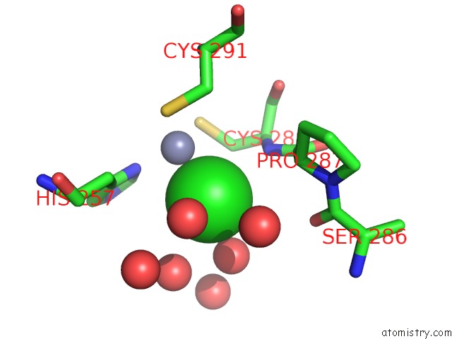
Mono view
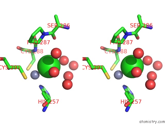
Stereo pair view
Mono view
Stereo pair view
A full contact list of Chlorine with other atoms in the Cl binding
site number 1 of Crystal Structure of A Soluble Variant of Full-Length Human APOBEC3G (pH 7.6) within 5.0Å range:
|
Reference:
A.Maiti,
W.Myint,
K.A.Delviks-Frankenberry,
S.Hou,
T.Kanai,
V.Balachandran,
C.Sierra Rodriguez,
R.Tripathi,
N.Kurt Yilmaz,
V.K.Pathak,
C.A.Schiffer,
H.Matsuo.
Crystal Structure of A Soluble APOBEC3G Variant Suggests Ssdna to Bind in A Channel That Extends Between the Two Domains. J.Mol.Biol. V. 432 6042 2020.
ISSN: ESSN 1089-8638
PubMed: 33098858
DOI: 10.1016/J.JMB.2020.10.020
Page generated: Sat Jul 12 21:18:34 2025
ISSN: ESSN 1089-8638
PubMed: 33098858
DOI: 10.1016/J.JMB.2020.10.020
Last articles
Fe in 2YXOFe in 2YRS
Fe in 2YXC
Fe in 2YNM
Fe in 2YVJ
Fe in 2YP1
Fe in 2YU2
Fe in 2YU1
Fe in 2YQB
Fe in 2YOO