Chlorine »
PDB 6x5s-6xi7 »
6xg5 »
Chlorine in PDB 6xg5: X-Ray Structure of Escherichia Coli Dihydrofolate Reductase in Complex with Trimethoprim
Enzymatic activity of X-Ray Structure of Escherichia Coli Dihydrofolate Reductase in Complex with Trimethoprim
All present enzymatic activity of X-Ray Structure of Escherichia Coli Dihydrofolate Reductase in Complex with Trimethoprim:
1.5.1.3;
1.5.1.3;
Protein crystallography data
The structure of X-Ray Structure of Escherichia Coli Dihydrofolate Reductase in Complex with Trimethoprim, PDB code: 6xg5
was solved by
I.K.Gaszek,
M.S.Manna,
D.Borek,
E.Toprak,
with X-Ray Crystallography technique. A brief refinement statistics is given in the table below:
| Resolution Low / High (Å) | 47.70 / 1.90 |
| Space group | P 32 2 1 |
| Cell size a, b, c (Å), α, β, γ (°) | 61.809, 61.809, 104.63, 90, 90, 120 |
| R / Rfree (%) | 23.6 / 26.3 |
Chlorine Binding Sites:
The binding sites of Chlorine atom in the X-Ray Structure of Escherichia Coli Dihydrofolate Reductase in Complex with Trimethoprim
(pdb code 6xg5). This binding sites where shown within
5.0 Angstroms radius around Chlorine atom.
In total only one binding site of Chlorine was determined in the X-Ray Structure of Escherichia Coli Dihydrofolate Reductase in Complex with Trimethoprim, PDB code: 6xg5:
In total only one binding site of Chlorine was determined in the X-Ray Structure of Escherichia Coli Dihydrofolate Reductase in Complex with Trimethoprim, PDB code: 6xg5:
Chlorine binding site 1 out of 1 in 6xg5
Go back to
Chlorine binding site 1 out
of 1 in the X-Ray Structure of Escherichia Coli Dihydrofolate Reductase in Complex with Trimethoprim
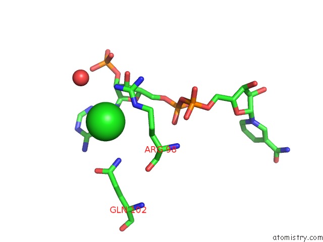
Mono view
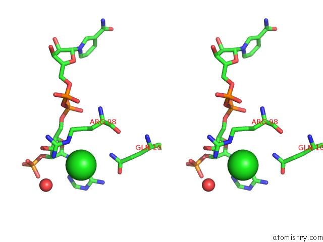
Stereo pair view
Mono view
Stereo pair view
A full contact list of Chlorine with other atoms in the Cl binding
site number 1 of X-Ray Structure of Escherichia Coli Dihydrofolate Reductase in Complex with Trimethoprim within 5.0Å range:
|
Reference:
M.S.Manna,
Y.T.Tamer,
I.Gaszek,
N.Poulides,
A.Ahmed,
F.C.R.Toprak,
D.Borek,
A.R.Atilgan,
J.Hulleman,
C.Atilgan,
U.Tambar,
E.Toprak.
Mutant Specific Drug Impedes the Evolution of Antibiotic Resistance in Escherichia Coli To Be Published.
Page generated: Mon Jul 29 17:11:27 2024
Last articles
Zn in 9J0NZn in 9J0O
Zn in 9J0P
Zn in 9FJX
Zn in 9EKB
Zn in 9C0F
Zn in 9CAH
Zn in 9CH0
Zn in 9CH3
Zn in 9CH1