Chlorine »
PDB 6xi9-6xqy »
6xi9 »
Chlorine in PDB 6xi9: X-Ray Crystal Structure of Mqne From Pedobacter Heparinus in Complex with Aminofutalosine and Methionine
Enzymatic activity of X-Ray Crystal Structure of Mqne From Pedobacter Heparinus in Complex with Aminofutalosine and Methionine
All present enzymatic activity of X-Ray Crystal Structure of Mqne From Pedobacter Heparinus in Complex with Aminofutalosine and Methionine:
2.5.1.120;
2.5.1.120;
Protein crystallography data
The structure of X-Ray Crystal Structure of Mqne From Pedobacter Heparinus in Complex with Aminofutalosine and Methionine, PDB code: 6xi9
was solved by
T.L.Grove,
J.B.Bonanno,
S.C.Almo,
with X-Ray Crystallography technique. A brief refinement statistics is given in the table below:
| Resolution Low / High (Å) | 19.96 / 2.14 |
| Space group | P 1 21 1 |
| Cell size a, b, c (Å), α, β, γ (°) | 72.512, 75.079, 83.269, 90.00, 107.13, 90.00 |
| R / Rfree (%) | 15.1 / 19.8 |
Other elements in 6xi9:
The structure of X-Ray Crystal Structure of Mqne From Pedobacter Heparinus in Complex with Aminofutalosine and Methionine also contains other interesting chemical elements:
| Iron | (Fe) | 8 atoms |
Chlorine Binding Sites:
The binding sites of Chlorine atom in the X-Ray Crystal Structure of Mqne From Pedobacter Heparinus in Complex with Aminofutalosine and Methionine
(pdb code 6xi9). This binding sites where shown within
5.0 Angstroms radius around Chlorine atom.
In total only one binding site of Chlorine was determined in the X-Ray Crystal Structure of Mqne From Pedobacter Heparinus in Complex with Aminofutalosine and Methionine, PDB code: 6xi9:
In total only one binding site of Chlorine was determined in the X-Ray Crystal Structure of Mqne From Pedobacter Heparinus in Complex with Aminofutalosine and Methionine, PDB code: 6xi9:
Chlorine binding site 1 out of 1 in 6xi9
Go back to
Chlorine binding site 1 out
of 1 in the X-Ray Crystal Structure of Mqne From Pedobacter Heparinus in Complex with Aminofutalosine and Methionine
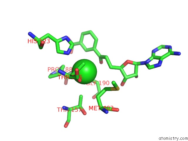
Mono view
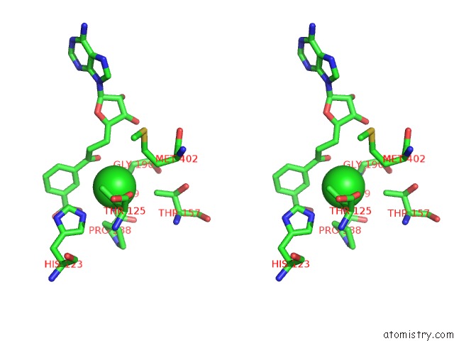
Stereo pair view
Mono view
Stereo pair view
A full contact list of Chlorine with other atoms in the Cl binding
site number 1 of X-Ray Crystal Structure of Mqne From Pedobacter Heparinus in Complex with Aminofutalosine and Methionine within 5.0Å range:
|
Reference:
A.G.Carl,
L.D.Harris,
M.Feng,
L.U.Nordstrom,
G.J.Gerfen,
G.B.Evans,
A.Silakov,
S.C.Almo,
T.L.Grove.
Narrow-Spectrum Antibiotic Targeting of the Radical Sam Enzyme Mqne in Menaquinone Biosynthesis. Biochemistry V. 59 2562 2020.
ISSN: ISSN 0006-2960
PubMed: 32627538
DOI: 10.1021/ACS.BIOCHEM.0C00070
Page generated: Mon Jul 29 17:15:17 2024
ISSN: ISSN 0006-2960
PubMed: 32627538
DOI: 10.1021/ACS.BIOCHEM.0C00070
Last articles
Zn in 9J0NZn in 9J0O
Zn in 9J0P
Zn in 9FJX
Zn in 9EKB
Zn in 9C0F
Zn in 9CAH
Zn in 9CH0
Zn in 9CH3
Zn in 9CH1