Chlorine »
PDB 6xi9-6xqy »
6xiz »
Chlorine in PDB 6xiz: Crystal Structure of Multi-Copper Oxidase From Pediococcus Acidilactici
Protein crystallography data
The structure of Crystal Structure of Multi-Copper Oxidase From Pediococcus Acidilactici, PDB code: 6xiz
was solved by
I.Pardo,
A.S.Soares,
R.Collins,
S.H.Partowmah,
E.A.Coler,
with X-Ray Crystallography technique. A brief refinement statistics is given in the table below:
| Resolution Low / High (Å) | 73.65 / 1.80 |
| Space group | P 1 21 1 |
| Cell size a, b, c (Å), α, β, γ (°) | 56.06, 147.3, 65.46, 90, 98.55, 90 |
| R / Rfree (%) | 11.4 / 16.1 |
Other elements in 6xiz:
The structure of Crystal Structure of Multi-Copper Oxidase From Pediococcus Acidilactici also contains other interesting chemical elements:
| Copper | (Cu) | 8 atoms |
Chlorine Binding Sites:
The binding sites of Chlorine atom in the Crystal Structure of Multi-Copper Oxidase From Pediococcus Acidilactici
(pdb code 6xiz). This binding sites where shown within
5.0 Angstroms radius around Chlorine atom.
In total 2 binding sites of Chlorine where determined in the Crystal Structure of Multi-Copper Oxidase From Pediococcus Acidilactici, PDB code: 6xiz:
Jump to Chlorine binding site number: 1; 2;
In total 2 binding sites of Chlorine where determined in the Crystal Structure of Multi-Copper Oxidase From Pediococcus Acidilactici, PDB code: 6xiz:
Jump to Chlorine binding site number: 1; 2;
Chlorine binding site 1 out of 2 in 6xiz
Go back to
Chlorine binding site 1 out
of 2 in the Crystal Structure of Multi-Copper Oxidase From Pediococcus Acidilactici
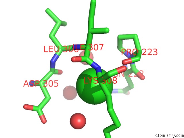
Mono view
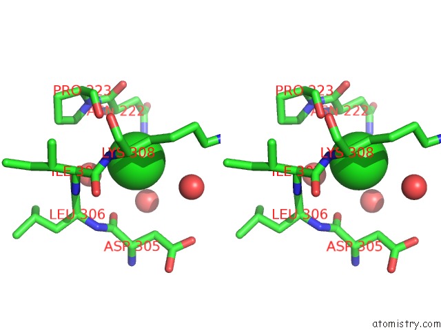
Stereo pair view
Mono view
Stereo pair view
A full contact list of Chlorine with other atoms in the Cl binding
site number 1 of Crystal Structure of Multi-Copper Oxidase From Pediococcus Acidilactici within 5.0Å range:
|
Chlorine binding site 2 out of 2 in 6xiz
Go back to
Chlorine binding site 2 out
of 2 in the Crystal Structure of Multi-Copper Oxidase From Pediococcus Acidilactici
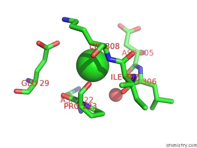
Mono view
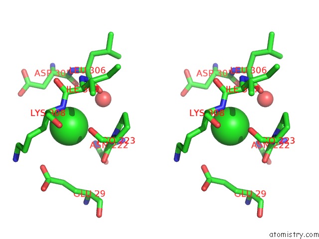
Stereo pair view
Mono view
Stereo pair view
A full contact list of Chlorine with other atoms in the Cl binding
site number 2 of Crystal Structure of Multi-Copper Oxidase From Pediococcus Acidilactici within 5.0Å range:
|
Reference:
I.Olmeda,
P.Casino,
R.E.Collins,
R.Sendra,
S.Callejon,
J.Huesa,
A.S.Soares,
S.Ferrer,
I.Pardo.
Structural Analysis and Biochemical Properties of Laccase Enzymes From Two Pediococcus Species. Microb Biotechnol 2021.
ISSN: ISSN 1751-7915
PubMed: 33635570
DOI: 10.1111/1751-7915.13751
Page generated: Mon Jul 29 17:15:17 2024
ISSN: ISSN 1751-7915
PubMed: 33635570
DOI: 10.1111/1751-7915.13751
Last articles
Zn in 9J0NZn in 9J0O
Zn in 9J0P
Zn in 9FJX
Zn in 9EKB
Zn in 9C0F
Zn in 9CAH
Zn in 9CH0
Zn in 9CH3
Zn in 9CH1