Chlorine »
PDB 6xi9-6xqy »
6xqk »
Chlorine in PDB 6xqk: Crystal Structure of the D/D Domain of Pka From S. Cerevisiae
Protein crystallography data
The structure of Crystal Structure of the D/D Domain of Pka From S. Cerevisiae, PDB code: 6xqk
was solved by
N.Larrieux,
N.Gonzalez Bardeci,
F.Trajtenberg,
A.Buschiazzo,
with X-Ray Crystallography technique. A brief refinement statistics is given in the table below:
| Resolution Low / High (Å) | 64.80 / 2.56 |
| Space group | P 1 21 1 |
| Cell size a, b, c (Å), α, β, γ (°) | 56.824, 52.134, 68.832, 90, 109.7, 90 |
| R / Rfree (%) | 21.4 / 27 |
Chlorine Binding Sites:
The binding sites of Chlorine atom in the Crystal Structure of the D/D Domain of Pka From S. Cerevisiae
(pdb code 6xqk). This binding sites where shown within
5.0 Angstroms radius around Chlorine atom.
In total only one binding site of Chlorine was determined in the Crystal Structure of the D/D Domain of Pka From S. Cerevisiae, PDB code: 6xqk:
In total only one binding site of Chlorine was determined in the Crystal Structure of the D/D Domain of Pka From S. Cerevisiae, PDB code: 6xqk:
Chlorine binding site 1 out of 1 in 6xqk
Go back to
Chlorine binding site 1 out
of 1 in the Crystal Structure of the D/D Domain of Pka From S. Cerevisiae
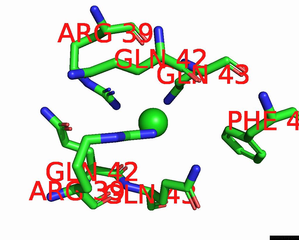
Mono view
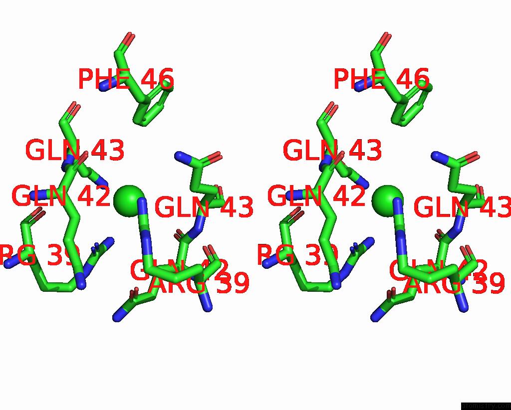
Stereo pair view
Mono view
Stereo pair view
A full contact list of Chlorine with other atoms in the Cl binding
site number 1 of Crystal Structure of the D/D Domain of Pka From S. Cerevisiae within 5.0Å range:
|
Reference:
N.Gonzalez Bardeci,
E.Tofolon,
F.Trajtenberg,
J.Caramelo,
N.Larrieux,
S.Rossi,
A.Buschiazzo,
S.Moreno.
The Crystal Structure of Yeast Regulatory Subunit Reveals Key Evolutionary Insights Into Protein Kinase A Oligomerization J.Struct.Biol. 2021.
ISSN: ESSN 1095-8657
DOI: 10.1016/J.JSB.2021.107732
Page generated: Mon Jul 29 17:19:44 2024
ISSN: ESSN 1095-8657
DOI: 10.1016/J.JSB.2021.107732
Last articles
Zn in 9J0NZn in 9J0O
Zn in 9J0P
Zn in 9FJX
Zn in 9EKB
Zn in 9C0F
Zn in 9CAH
Zn in 9CH0
Zn in 9CH3
Zn in 9CH1