Chlorine »
PDB 6xqz-6xys »
6xyb »
Chlorine in PDB 6xyb: Crystal Structure of Q4D6Q6, A Conserved Kinetoplastid-Specific Protein From Trypanosoma Cruzi
Protein crystallography data
The structure of Crystal Structure of Q4D6Q6, A Conserved Kinetoplastid-Specific Protein From Trypanosoma Cruzi, PDB code: 6xyb
was solved by
Y.Roske,
U.Heinemann,
with X-Ray Crystallography technique. A brief refinement statistics is given in the table below:
| Resolution Low / High (Å) | 34.71 / 1.47 |
| Space group | P 1 21 1 |
| Cell size a, b, c (Å), α, β, γ (°) | 66.701, 44.487, 69.544, 90.00, 93.46, 90.00 |
| R / Rfree (%) | 14.3 / 18.6 |
Other elements in 6xyb:
The structure of Crystal Structure of Q4D6Q6, A Conserved Kinetoplastid-Specific Protein From Trypanosoma Cruzi also contains other interesting chemical elements:
| Magnesium | (Mg) | 2 atoms |
| Potassium | (K) | 1 atom |
| Iodine | (I) | 2 atoms |
Chlorine Binding Sites:
The binding sites of Chlorine atom in the Crystal Structure of Q4D6Q6, A Conserved Kinetoplastid-Specific Protein From Trypanosoma Cruzi
(pdb code 6xyb). This binding sites where shown within
5.0 Angstroms radius around Chlorine atom.
In total only one binding site of Chlorine was determined in the Crystal Structure of Q4D6Q6, A Conserved Kinetoplastid-Specific Protein From Trypanosoma Cruzi, PDB code: 6xyb:
In total only one binding site of Chlorine was determined in the Crystal Structure of Q4D6Q6, A Conserved Kinetoplastid-Specific Protein From Trypanosoma Cruzi, PDB code: 6xyb:
Chlorine binding site 1 out of 1 in 6xyb
Go back to
Chlorine binding site 1 out
of 1 in the Crystal Structure of Q4D6Q6, A Conserved Kinetoplastid-Specific Protein From Trypanosoma Cruzi
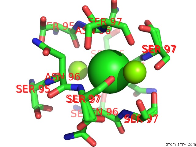
Mono view
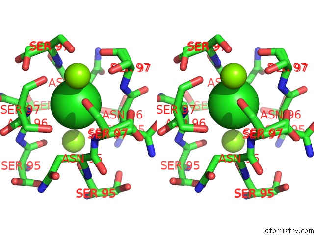
Stereo pair view
Mono view
Stereo pair view
A full contact list of Chlorine with other atoms in the Cl binding
site number 1 of Crystal Structure of Q4D6Q6, A Conserved Kinetoplastid-Specific Protein From Trypanosoma Cruzi within 5.0Å range:
|
Reference:
E.Dias D'andrea,
Y.Roske,
G.A.P.De Oliveira,
N.Cremer,
A.Diehl,
P.Schmieder,
U.Heinemann,
H.Oschkinat,
J.Ricardo Pires.
Crystal Structure of Q4D6Q6, A Conserved Kinetoplastid-Specific Protein From Trypanosoma Cruzi. J.Struct.Biol. 07536 2020.
ISSN: ESSN 1095-8657
PubMed: 32473201
DOI: 10.1016/J.JSB.2020.107536
Page generated: Mon Jul 29 17:26:17 2024
ISSN: ESSN 1095-8657
PubMed: 32473201
DOI: 10.1016/J.JSB.2020.107536
Last articles
Zn in 9J0NZn in 9J0O
Zn in 9J0P
Zn in 9FJX
Zn in 9EKB
Zn in 9C0F
Zn in 9CAH
Zn in 9CH0
Zn in 9CH3
Zn in 9CH1