Chlorine »
PDB 6xys-6y7z »
6y3u »
Chlorine in PDB 6y3u: Crystal Structure of Ppargamma in Complex with Compound (R)-16
Protein crystallography data
The structure of Crystal Structure of Ppargamma in Complex with Compound (R)-16, PDB code: 6y3u
was solved by
A.Chaikuad,
T.Hanke,
C.H.Arrowsmith,
A.M.Edwards,
C.Bountra,
D.Merk,
S.Knapp,
Structural Genomics Consortium (Sgc),
with X-Ray Crystallography technique. A brief refinement statistics is given in the table below:
| Resolution Low / High (Å) | 43.88 / 2.62 |
| Space group | P 41 21 2 |
| Cell size a, b, c (Å), α, β, γ (°) | 62.059, 62.059, 167.021, 90.00, 90.00, 90.00 |
| R / Rfree (%) | 20.6 / 25.4 |
Chlorine Binding Sites:
The binding sites of Chlorine atom in the Crystal Structure of Ppargamma in Complex with Compound (R)-16
(pdb code 6y3u). This binding sites where shown within
5.0 Angstroms radius around Chlorine atom.
In total 2 binding sites of Chlorine where determined in the Crystal Structure of Ppargamma in Complex with Compound (R)-16, PDB code: 6y3u:
Jump to Chlorine binding site number: 1; 2;
In total 2 binding sites of Chlorine where determined in the Crystal Structure of Ppargamma in Complex with Compound (R)-16, PDB code: 6y3u:
Jump to Chlorine binding site number: 1; 2;
Chlorine binding site 1 out of 2 in 6y3u
Go back to
Chlorine binding site 1 out
of 2 in the Crystal Structure of Ppargamma in Complex with Compound (R)-16
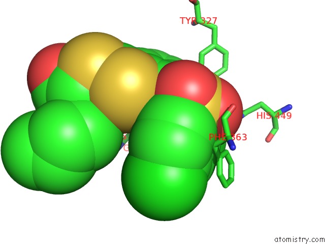
Mono view
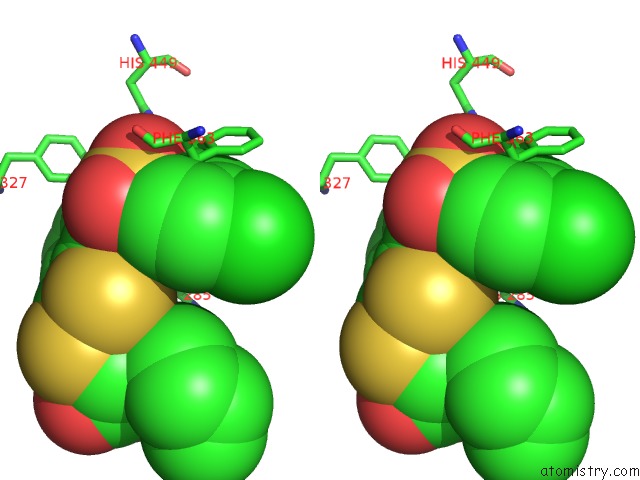
Stereo pair view
Mono view
Stereo pair view
A full contact list of Chlorine with other atoms in the Cl binding
site number 1 of Crystal Structure of Ppargamma in Complex with Compound (R)-16 within 5.0Å range:
|
Chlorine binding site 2 out of 2 in 6y3u
Go back to
Chlorine binding site 2 out
of 2 in the Crystal Structure of Ppargamma in Complex with Compound (R)-16
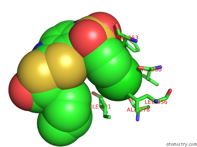
Mono view
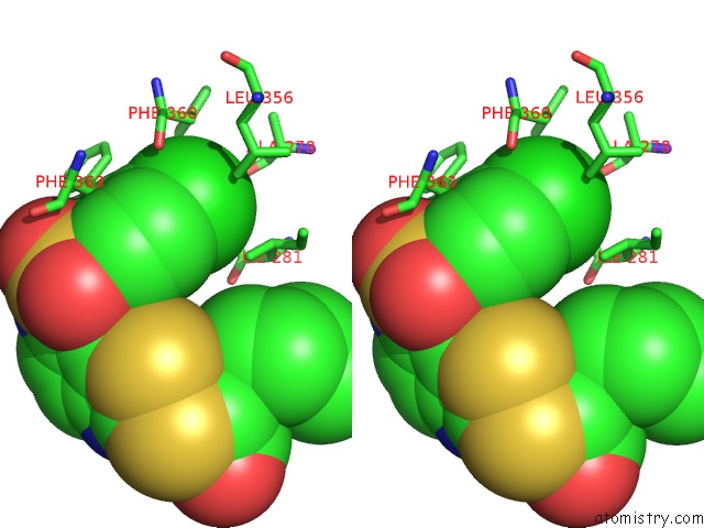
Stereo pair view
Mono view
Stereo pair view
A full contact list of Chlorine with other atoms in the Cl binding
site number 2 of Crystal Structure of Ppargamma in Complex with Compound (R)-16 within 5.0Å range:
|
Reference:
T.Hanke,
S.Y.Cheung,
W.Kilu,
J.Heering,
X.Ni,
V.Planz,
S.Schierle,
G.Faudone,
M.Friedrich,
M.Wanior,
O.Werz,
M.Windbergs,
E.Proschak,
M.Schubert-Zsilavecz,
A.Chaikuad,
S.Knapp,
D.Merk.
A Selective Modulator of Peroxisome Proliferator-Activated Receptor Gamma with Unprecedented Binding Mode. J.Med.Chem. 2020.
ISSN: ISSN 0022-2623
PubMed: 32267688
DOI: 10.1021/ACS.JMEDCHEM.9B01786
Page generated: Mon Jul 29 17:29:38 2024
ISSN: ISSN 0022-2623
PubMed: 32267688
DOI: 10.1021/ACS.JMEDCHEM.9B01786
Last articles
Zn in 9JYWZn in 9IR4
Zn in 9IR3
Zn in 9GMX
Zn in 9GMW
Zn in 9JEJ
Zn in 9ERF
Zn in 9ERE
Zn in 9EGV
Zn in 9EGW