Chlorine »
PDB 6ynp-6yyc »
6ynq »
Chlorine in PDB 6ynq: Structure of Sars-Cov-2 Main Protease Bound to 2-Methyl-1-Tetralone.
Protein crystallography data
The structure of Structure of Sars-Cov-2 Main Protease Bound to 2-Methyl-1-Tetralone., PDB code: 6ynq
was solved by
S.Guenther,
P.Reinke,
D.Oberthuer,
O.Yefanov,
L.Gelisio,
H.Ginn,
J.Lieske,
M.Domaracky,
W.Brehm,
A.Rahmani Mashour,
T.A.White,
J.Knoska,
G.Penaesperanza,
F.Koua,
A.Tolstikova,
M.Groessler,
P.Fischer,
V.Hennicke,
H.Fleckenstein,
F.Trost,
M.Galchenkova,
Y.Gevorkov,
C.Li,
S.Awel,
L.X.Paulraj,
N.Ullah,
S.Falke,
B.Alves Franca,
M.Schwinzer,
H.Brognaro,
N.Werner,
M.Perbandt,
H.Tidow,
B.Seychell,
T.Beck,
S.Meier,
J.J.Doyle,
H.Giseler,
D.Melo,
I.Dunkel,
T.J.Lane,
A.Peck,
S.Saouane,
J.Hakanpaeae,
J.Meyer,
H.Noei,
P.Gribbon,
B.Ellinger,
M.Kuzikov,
M.Wolf,
L.Zhang,
C.Ehrt,
J.Pletzer-Zelgert,
J.Wollenhaupt,
C.Feiler,
M.Weiss,
E.C.Schulz,
P.Mehrabi,
B.Norton-Baker,
C.Schmidt,
K.Lorenzen,
R.Schubert,
H.Han,
A.Chari,
Y.Fernandez Garcia,
D.Turk,
R.Hilgenfeld,
M.Rarey,
A.Zaliani,
H.N.Chapman,
A.Pearson,
C.Betzel,
A.Meents,
with X-Ray Crystallography technique. A brief refinement statistics is given in the table below:
| Resolution Low / High (Å) | 27.55 / 1.80 |
| Space group | C 1 2 1 |
| Cell size a, b, c (Å), α, β, γ (°) | 113.083, 53.154, 44.650, 90.00, 102.96, 90.00 |
| R / Rfree (%) | 18.2 / 22.9 |
Chlorine Binding Sites:
The binding sites of Chlorine atom in the Structure of Sars-Cov-2 Main Protease Bound to 2-Methyl-1-Tetralone.
(pdb code 6ynq). This binding sites where shown within
5.0 Angstroms radius around Chlorine atom.
In total only one binding site of Chlorine was determined in the Structure of Sars-Cov-2 Main Protease Bound to 2-Methyl-1-Tetralone., PDB code: 6ynq:
In total only one binding site of Chlorine was determined in the Structure of Sars-Cov-2 Main Protease Bound to 2-Methyl-1-Tetralone., PDB code: 6ynq:
Chlorine binding site 1 out of 1 in 6ynq
Go back to
Chlorine binding site 1 out
of 1 in the Structure of Sars-Cov-2 Main Protease Bound to 2-Methyl-1-Tetralone.
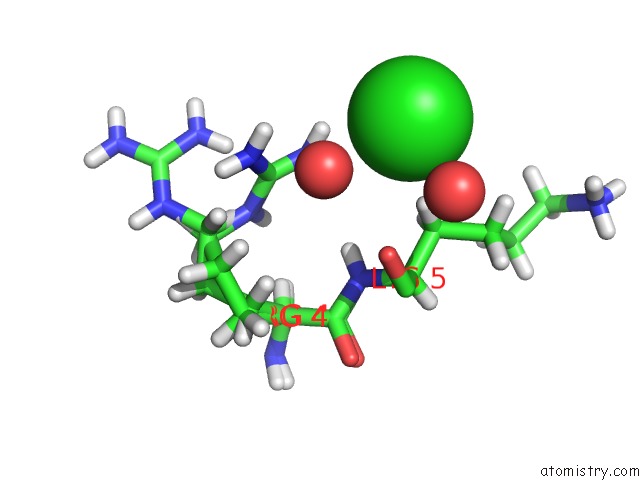
Mono view
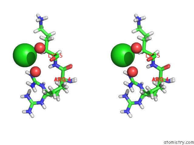
Stereo pair view
Mono view
Stereo pair view
A full contact list of Chlorine with other atoms in the Cl binding
site number 1 of Structure of Sars-Cov-2 Main Protease Bound to 2-Methyl-1-Tetralone. within 5.0Å range:
|
Reference:
S.Guenther,
P.Reinke,
D.Oberthuer,
O.Yefanov,
L.Gelisio,
H.Ginn,
J.Lieske,
W.Brehm,
A.Rahmani Mashour,
J.Knoska,
G.Pena Esperanza,
F.Koua,
A.Tolstikova,
M.Groessler,
H.Fleckenstein,
F.Trost,
M.Galchenkova,
Y.Gevorkov,
C.Li,
S.Awel,
L.X.Paulraj,
N.Ullah,
S.Falke,
B.Alves Franca,
M.Schwinzer,
H.Brognaro,
N.Werner,
M.Perbandt,
B.Seychell,
S.Meier,
H.Giseler,
D.Melo,
I.Dunkel,
T.J.Lane,
A.Peck,
S.Saouane,
J.Hakanpaeae,
J.Meyer,
H.Noei,
P.Gribbon,
B.Ellinger,
M.Kuzikov,
M.Wolf,
L.Zhang,
C.Ehrt,
J.Pletzer-Zelgert,
J.Wollenhaupt,
C.Feiler,
M.Weiss,
E.C.Schulz,
P.Mehrabi,
B.Norton-Baker,
C.Schmidt,
K.Lorenzen,
R.Schubert,
H.Han,
A.Chari,
Y.Fernandez Garcia,
R.Hilgenfeld,
M.Rarey,
A.Zaliani,
H.N.Chapman,
A.Pearson,
C.Betzel,
A.Meents.
Structure of Sars-Cov-2 Main Protease Bound 2-Methyl-1-Tetralone. To Be Published.
Page generated: Sat Jul 12 22:09:56 2025
Last articles
F in 7NZNF in 7NXN
F in 7NWB
F in 7NXS
F in 7NXY
F in 7NWC
F in 7NXO
F in 7NWM
F in 7NWE
F in 7NWK