Chlorine »
PDB 6yyc-6zbo »
6yyf »
Chlorine in PDB 6yyf: Crystal Structure of 5-Chloroindoline-2,3-Dione Covalently Bound to the pH Domain of Bruton'S Tyrosine Kinase Mutant R28C
Enzymatic activity of Crystal Structure of 5-Chloroindoline-2,3-Dione Covalently Bound to the pH Domain of Bruton'S Tyrosine Kinase Mutant R28C
All present enzymatic activity of Crystal Structure of 5-Chloroindoline-2,3-Dione Covalently Bound to the pH Domain of Bruton'S Tyrosine Kinase Mutant R28C:
2.7.10.2;
2.7.10.2;
Protein crystallography data
The structure of Crystal Structure of 5-Chloroindoline-2,3-Dione Covalently Bound to the pH Domain of Bruton'S Tyrosine Kinase Mutant R28C, PDB code: 6yyf
was solved by
P.Brear,
J.Wagstaff,
M.Hyvonen,
with X-Ray Crystallography technique. A brief refinement statistics is given in the table below:
| Resolution Low / High (Å) | 36.74 / 1.93 |
| Space group | P 1 21 1 |
| Cell size a, b, c (Å), α, β, γ (°) | 46.9, 60.29, 57.4, 90, 98.93, 90 |
| R / Rfree (%) | 19 / 24.9 |
Other elements in 6yyf:
The structure of Crystal Structure of 5-Chloroindoline-2,3-Dione Covalently Bound to the pH Domain of Bruton'S Tyrosine Kinase Mutant R28C also contains other interesting chemical elements:
| Zinc | (Zn) | 2 atoms |
| Magnesium | (Mg) | 2 atoms |
Chlorine Binding Sites:
The binding sites of Chlorine atom in the Crystal Structure of 5-Chloroindoline-2,3-Dione Covalently Bound to the pH Domain of Bruton'S Tyrosine Kinase Mutant R28C
(pdb code 6yyf). This binding sites where shown within
5.0 Angstroms radius around Chlorine atom.
In total 2 binding sites of Chlorine where determined in the Crystal Structure of 5-Chloroindoline-2,3-Dione Covalently Bound to the pH Domain of Bruton'S Tyrosine Kinase Mutant R28C, PDB code: 6yyf:
Jump to Chlorine binding site number: 1; 2;
In total 2 binding sites of Chlorine where determined in the Crystal Structure of 5-Chloroindoline-2,3-Dione Covalently Bound to the pH Domain of Bruton'S Tyrosine Kinase Mutant R28C, PDB code: 6yyf:
Jump to Chlorine binding site number: 1; 2;
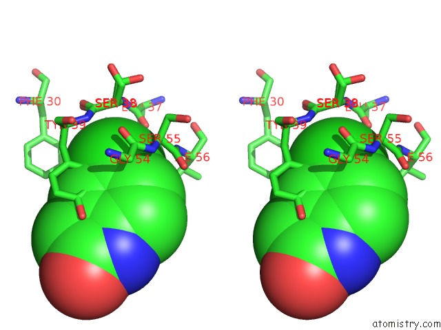
Chlorine binding site 1 out of 2 in 6yyf
Go back to
Chlorine binding site 1 out
of 2 in the Crystal Structure of 5-Chloroindoline-2,3-Dione Covalently Bound to the pH Domain of Bruton'S Tyrosine Kinase Mutant R28C

Mono view

Stereo pair view
Mono view
Stereo pair view
A full contact list of Chlorine with other atoms in the Cl binding
site number 1 of Crystal Structure of 5-Chloroindoline-2,3-Dione Covalently Bound to the pH Domain of Bruton'S Tyrosine Kinase Mutant R28C within 5.0Å range:
|
Chlorine binding site 2 out of 2 in 6yyf
Go back to
Chlorine binding site 2 out
of 2 in the Crystal Structure of 5-Chloroindoline-2,3-Dione Covalently Bound to the pH Domain of Bruton'S Tyrosine Kinase Mutant R28C

Mono view
Stereo pair view
Mono view
Stereo pair view
A full contact list of Chlorine with other atoms in the Cl binding
site number 2 of Crystal Structure of 5-Chloroindoline-2,3-Dione Covalently Bound to the pH Domain of Bruton'S Tyrosine Kinase Mutant R28C within 5.0Å range:
|
Reference:
P.Brear,
G.Fischer,
M.May,
T.Pantelejevs,
R.Mathieu,
M.Rossmann,
J.Wagstaff,
B.Blaszczyk,
M.Hyvonen.
Optimising Crystallographic Systems For Structure-Guided Drug Discovery To Be Published.
Page generated: Mon Jul 29 17:55:13 2024
Last articles
Ca in 5PAJCa in 5PAI
Ca in 5PAG
Ca in 5PAF
Ca in 5P2P
Ca in 5PAC
Ca in 5PAE
Ca in 5PAB
Ca in 5PA8
Ca in 5PA9