Chlorine »
PDB 7b1o-7bab »
7b2c »
Chlorine in PDB 7b2c: Crystal Structure of the Ethyl-Coenzyme M Reductase From Candidatus Ethanoperedens Thermophilum Gassed with Xenon
Enzymatic activity of Crystal Structure of the Ethyl-Coenzyme M Reductase From Candidatus Ethanoperedens Thermophilum Gassed with Xenon
All present enzymatic activity of Crystal Structure of the Ethyl-Coenzyme M Reductase From Candidatus Ethanoperedens Thermophilum Gassed with Xenon:
2.8.4.1;
2.8.4.1;
Protein crystallography data
The structure of Crystal Structure of the Ethyl-Coenzyme M Reductase From Candidatus Ethanoperedens Thermophilum Gassed with Xenon, PDB code: 7b2c
was solved by
T.Wagner,
O.N.Lemaire,
S.Engilberge,
with X-Ray Crystallography technique. A brief refinement statistics is given in the table below:
| Resolution Low / High (Å) | 35.90 / 1.80 |
| Space group | P 1 21 1 |
| Cell size a, b, c (Å), α, β, γ (°) | 83.791, 147.186, 113.376, 90, 107.2, 90 |
| R / Rfree (%) | 17.4 / 20.1 |
Other elements in 7b2c:
The structure of Crystal Structure of the Ethyl-Coenzyme M Reductase From Candidatus Ethanoperedens Thermophilum Gassed with Xenon also contains other interesting chemical elements:
| Magnesium | (Mg) | 1 atom |
| Potassium | (K) | 17 atoms |
| Nickel | (Ni) | 2 atoms |
| Xenon | (Xe) | 16 atoms |
| Sodium | (Na) | 5 atoms |
Chlorine Binding Sites:
The binding sites of Chlorine atom in the Crystal Structure of the Ethyl-Coenzyme M Reductase From Candidatus Ethanoperedens Thermophilum Gassed with Xenon
(pdb code 7b2c). This binding sites where shown within
5.0 Angstroms radius around Chlorine atom.
In total 5 binding sites of Chlorine where determined in the Crystal Structure of the Ethyl-Coenzyme M Reductase From Candidatus Ethanoperedens Thermophilum Gassed with Xenon, PDB code: 7b2c:
Jump to Chlorine binding site number: 1; 2; 3; 4; 5;
In total 5 binding sites of Chlorine where determined in the Crystal Structure of the Ethyl-Coenzyme M Reductase From Candidatus Ethanoperedens Thermophilum Gassed with Xenon, PDB code: 7b2c:
Jump to Chlorine binding site number: 1; 2; 3; 4; 5;
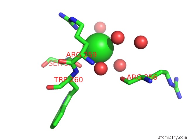
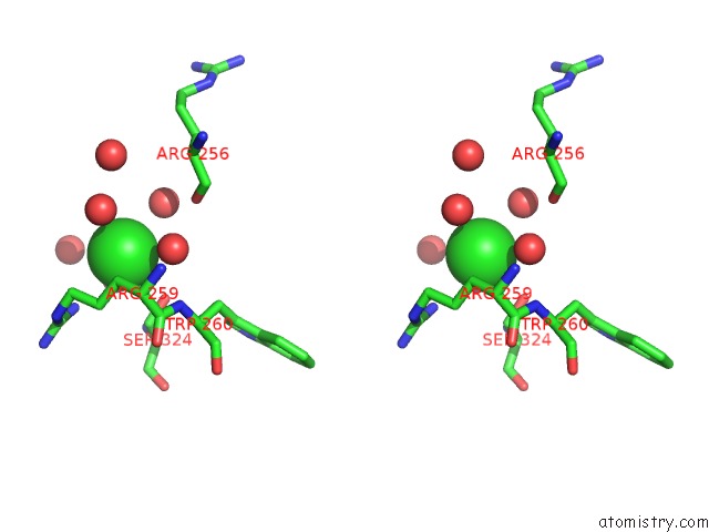
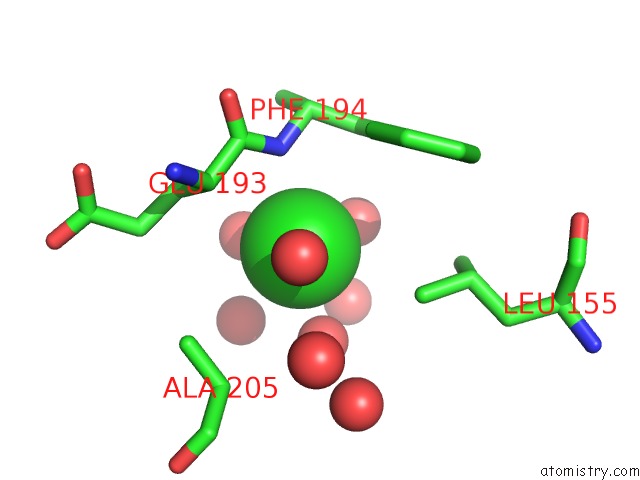
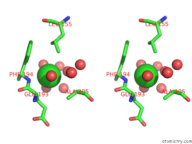
Chlorine binding site 1 out of 5 in 7b2c
Go back to
Chlorine binding site 1 out
of 5 in the Crystal Structure of the Ethyl-Coenzyme M Reductase From Candidatus Ethanoperedens Thermophilum Gassed with Xenon
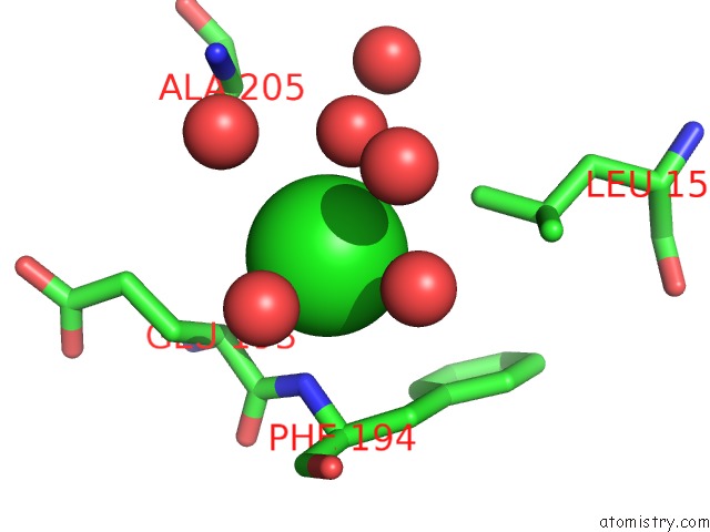
Mono view
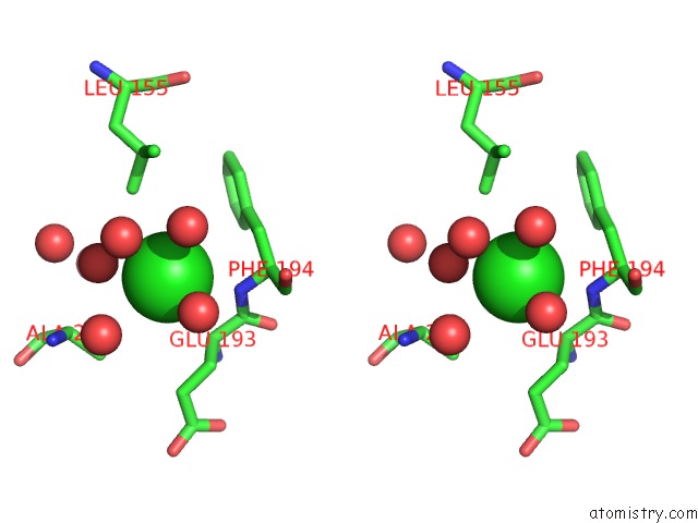
Stereo pair view
Mono view
Stereo pair view
A full contact list of Chlorine with other atoms in the Cl binding
site number 1 of Crystal Structure of the Ethyl-Coenzyme M Reductase From Candidatus Ethanoperedens Thermophilum Gassed with Xenon within 5.0Å range:
|
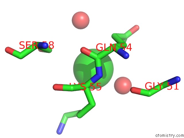
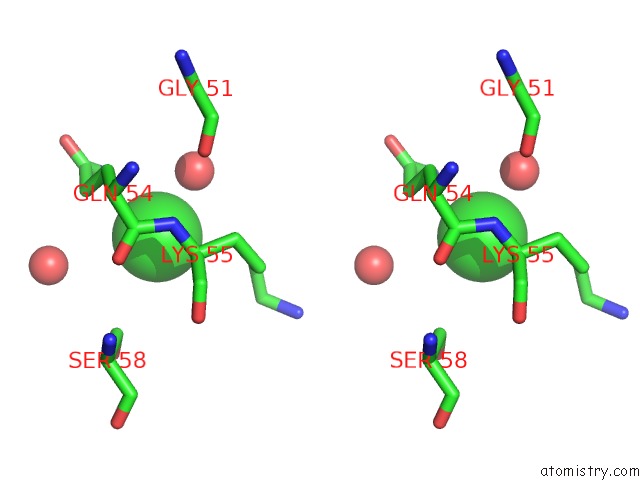
Chlorine binding site 2 out of 5 in 7b2c
Go back to
Chlorine binding site 2 out
of 5 in the Crystal Structure of the Ethyl-Coenzyme M Reductase From Candidatus Ethanoperedens Thermophilum Gassed with Xenon
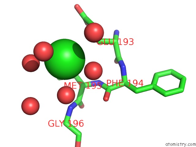
Mono view
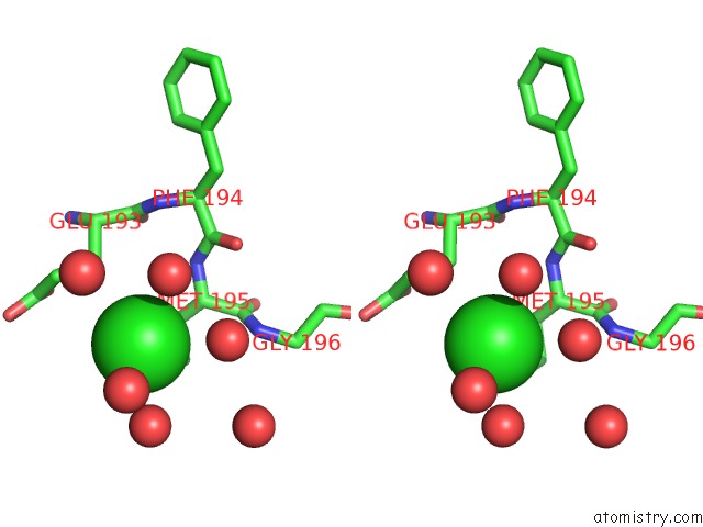
Stereo pair view
Mono view
Stereo pair view
A full contact list of Chlorine with other atoms in the Cl binding
site number 2 of Crystal Structure of the Ethyl-Coenzyme M Reductase From Candidatus Ethanoperedens Thermophilum Gassed with Xenon within 5.0Å range:
|
Chlorine binding site 3 out of 5 in 7b2c
Go back to
Chlorine binding site 3 out
of 5 in the Crystal Structure of the Ethyl-Coenzyme M Reductase From Candidatus Ethanoperedens Thermophilum Gassed with Xenon
Mono view
Stereo pair view
Mono view
Stereo pair view
A full contact list of Chlorine with other atoms in the Cl binding
site number 3 of Crystal Structure of the Ethyl-Coenzyme M Reductase From Candidatus Ethanoperedens Thermophilum Gassed with Xenon within 5.0Å range:
|
Chlorine binding site 4 out of 5 in 7b2c
Go back to
Chlorine binding site 4 out
of 5 in the Crystal Structure of the Ethyl-Coenzyme M Reductase From Candidatus Ethanoperedens Thermophilum Gassed with Xenon
Mono view
Stereo pair view
Mono view
Stereo pair view
A full contact list of Chlorine with other atoms in the Cl binding
site number 4 of Crystal Structure of the Ethyl-Coenzyme M Reductase From Candidatus Ethanoperedens Thermophilum Gassed with Xenon within 5.0Å range:
|
Chlorine binding site 5 out of 5 in 7b2c
Go back to
Chlorine binding site 5 out
of 5 in the Crystal Structure of the Ethyl-Coenzyme M Reductase From Candidatus Ethanoperedens Thermophilum Gassed with Xenon
Mono view
Stereo pair view
Mono view
Stereo pair view
A full contact list of Chlorine with other atoms in the Cl binding
site number 5 of Crystal Structure of the Ethyl-Coenzyme M Reductase From Candidatus Ethanoperedens Thermophilum Gassed with Xenon within 5.0Å range:
|
Reference:
C.J.Hahn,
O.N.Lemaire,
J.Kahnt,
S.Engilberge,
G.Wegener,
T.Wagner.
Crystal Structure of A Key Enzyme For Anaerobic Ethane Activation. Science V. 373 118 2021.
ISSN: ESSN 1095-9203
PubMed: 34210888
DOI: 10.1126/SCIENCE.ABG1765
Page generated: Mon Jul 29 18:57:55 2024
ISSN: ESSN 1095-9203
PubMed: 34210888
DOI: 10.1126/SCIENCE.ABG1765
Last articles
Cl in 2VOICl in 2VOB
Cl in 2VO6
Cl in 2VO7
Cl in 2VOF
Cl in 2VO5
Cl in 2VMF
Cl in 2VNT
Cl in 2VO3
Cl in 2VO0