Chlorine »
PDB 7c74-7ckz »
7cjz »
Chlorine in PDB 7cjz: Room Temperature Structure of Lysozyme Delivered in Lard By Serial Millisecond Crystallography
Enzymatic activity of Room Temperature Structure of Lysozyme Delivered in Lard By Serial Millisecond Crystallography
All present enzymatic activity of Room Temperature Structure of Lysozyme Delivered in Lard By Serial Millisecond Crystallography:
3.2.1.17;
3.2.1.17;
Protein crystallography data
The structure of Room Temperature Structure of Lysozyme Delivered in Lard By Serial Millisecond Crystallography, PDB code: 7cjz
was solved by
K.H.Nam,
with X-Ray Crystallography technique. A brief refinement statistics is given in the table below:
| Resolution Low / High (Å) | 56.18 / 1.75 |
| Space group | P 43 21 2 |
| Cell size a, b, c (Å), α, β, γ (°) | 79.450, 79.450, 38.470, 90.00, 90.00, 90.00 |
| R / Rfree (%) | 16.5 / 18.7 |
Other elements in 7cjz:
The structure of Room Temperature Structure of Lysozyme Delivered in Lard By Serial Millisecond Crystallography also contains other interesting chemical elements:
| Sodium | (Na) | 2 atoms |
Chlorine Binding Sites:
The binding sites of Chlorine atom in the Room Temperature Structure of Lysozyme Delivered in Lard By Serial Millisecond Crystallography
(pdb code 7cjz). This binding sites where shown within
5.0 Angstroms radius around Chlorine atom.
In total 2 binding sites of Chlorine where determined in the Room Temperature Structure of Lysozyme Delivered in Lard By Serial Millisecond Crystallography, PDB code: 7cjz:
Jump to Chlorine binding site number: 1; 2;
In total 2 binding sites of Chlorine where determined in the Room Temperature Structure of Lysozyme Delivered in Lard By Serial Millisecond Crystallography, PDB code: 7cjz:
Jump to Chlorine binding site number: 1; 2;
Chlorine binding site 1 out of 2 in 7cjz
Go back to
Chlorine binding site 1 out
of 2 in the Room Temperature Structure of Lysozyme Delivered in Lard By Serial Millisecond Crystallography

Mono view
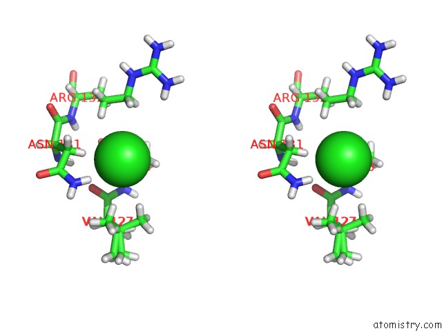
Stereo pair view
Mono view
Stereo pair view
A full contact list of Chlorine with other atoms in the Cl binding
site number 1 of Room Temperature Structure of Lysozyme Delivered in Lard By Serial Millisecond Crystallography within 5.0Å range:
|
Chlorine binding site 2 out of 2 in 7cjz
Go back to
Chlorine binding site 2 out
of 2 in the Room Temperature Structure of Lysozyme Delivered in Lard By Serial Millisecond Crystallography

Mono view
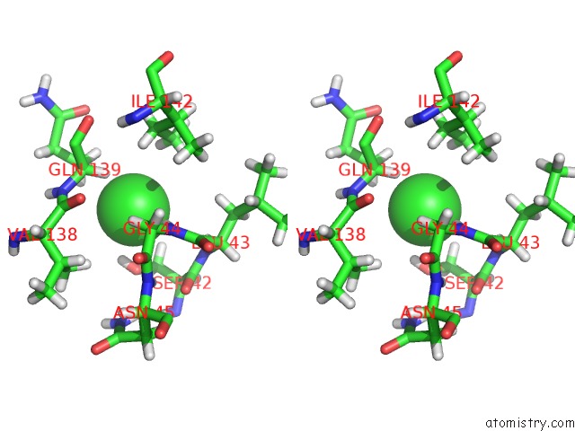
Stereo pair view
Mono view
Stereo pair view
A full contact list of Chlorine with other atoms in the Cl binding
site number 2 of Room Temperature Structure of Lysozyme Delivered in Lard By Serial Millisecond Crystallography within 5.0Å range:
|
Reference:
K.H.Nam,
K.H.Nam.
N/A N/A.
ISSN: ESSN 1422-0067
PubMed: 32825186
DOI: 10.3390/IJMS21175977
Page generated: Mon Jul 29 19:42:26 2024
ISSN: ESSN 1422-0067
PubMed: 32825186
DOI: 10.3390/IJMS21175977
Last articles
Zn in 9J0NZn in 9J0O
Zn in 9J0P
Zn in 9FJX
Zn in 9EKB
Zn in 9C0F
Zn in 9CAH
Zn in 9CH0
Zn in 9CH3
Zn in 9CH1