Chlorine »
PDB 7cvt-7daf »
7cwi »
Chlorine in PDB 7cwi: Crystal Structure of Beta-Galactosidase II From Bacillus Circulans
Protein crystallography data
The structure of Crystal Structure of Beta-Galactosidase II From Bacillus Circulans, PDB code: 7cwi
was solved by
H.Hong,
H.Seo,
with X-Ray Crystallography technique. A brief refinement statistics is given in the table below:
| Resolution Low / High (Å) | 31.77 / 1.95 |
| Space group | P 32 2 1 |
| Cell size a, b, c (Å), α, β, γ (°) | 159.802, 159.802, 96.167, 90.00, 90.00, 120.00 |
| R / Rfree (%) | 15.6 / 18.7 |
Chlorine Binding Sites:
The binding sites of Chlorine atom in the Crystal Structure of Beta-Galactosidase II From Bacillus Circulans
(pdb code 7cwi). This binding sites where shown within
5.0 Angstroms radius around Chlorine atom.
In total only one binding site of Chlorine was determined in the Crystal Structure of Beta-Galactosidase II From Bacillus Circulans, PDB code: 7cwi:
In total only one binding site of Chlorine was determined in the Crystal Structure of Beta-Galactosidase II From Bacillus Circulans, PDB code: 7cwi:
Chlorine binding site 1 out of 1 in 7cwi
Go back to
Chlorine binding site 1 out
of 1 in the Crystal Structure of Beta-Galactosidase II From Bacillus Circulans

Mono view
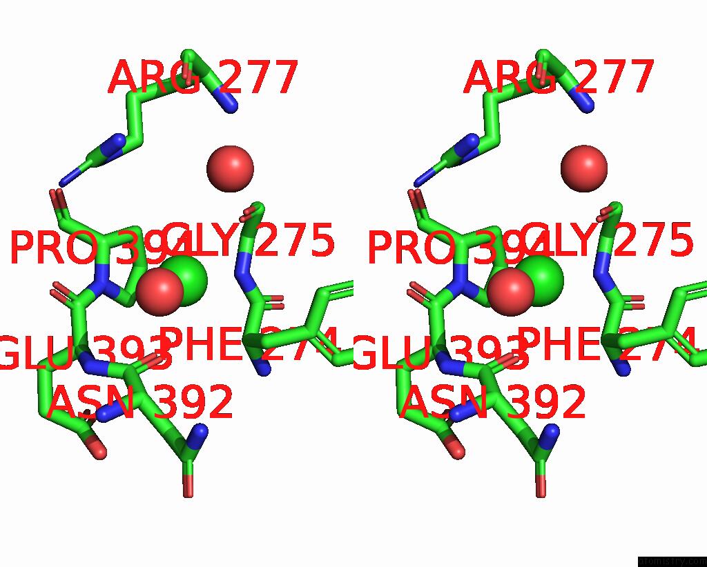
Stereo pair view
Mono view
Stereo pair view
A full contact list of Chlorine with other atoms in the Cl binding
site number 1 of Crystal Structure of Beta-Galactosidase II From Bacillus Circulans within 5.0Å range:
|
Reference:
J.Y.Choi,
H.Hong,
H.Seo,
J.G.Pan,
E.J.Kim,
P.J.Maeng,
T.H.Yang,
K.J.Kim.
High Galacto-Oligosaccharide Production and A Structural Model For Transgalactosylation of Beta-Galactosidase II From Bacillus Circulans . J.Agric.Food Chem. V. 68 13806 2020.
ISSN: ESSN 1520-5118
PubMed: 33169609
DOI: 10.1021/ACS.JAFC.0C05871
Page generated: Sat Jul 12 23:54:25 2025
ISSN: ESSN 1520-5118
PubMed: 33169609
DOI: 10.1021/ACS.JAFC.0C05871
Last articles
Fe in 2YXOFe in 2YRS
Fe in 2YXC
Fe in 2YNM
Fe in 2YVJ
Fe in 2YP1
Fe in 2YU2
Fe in 2YU1
Fe in 2YQB
Fe in 2YOO