Chlorine »
PDB 7daf-7dq0 »
7dfn »
Chlorine in PDB 7dfn: Crystal Structure of Glycoside Hydrolase Family 11 Beta-Xylanase From Streptomyces Olivaceoviridis E-86 in Complex with Alpha-L- Arabinofuranosyl Xylotetraose
Enzymatic activity of Crystal Structure of Glycoside Hydrolase Family 11 Beta-Xylanase From Streptomyces Olivaceoviridis E-86 in Complex with Alpha-L- Arabinofuranosyl Xylotetraose
All present enzymatic activity of Crystal Structure of Glycoside Hydrolase Family 11 Beta-Xylanase From Streptomyces Olivaceoviridis E-86 in Complex with Alpha-L- Arabinofuranosyl Xylotetraose:
3.2.1.8;
3.2.1.8;
Protein crystallography data
The structure of Crystal Structure of Glycoside Hydrolase Family 11 Beta-Xylanase From Streptomyces Olivaceoviridis E-86 in Complex with Alpha-L- Arabinofuranosyl Xylotetraose, PDB code: 7dfn
was solved by
Z.Fujimoto,
N.Kishine,
S.Kaneko,
with X-Ray Crystallography technique. A brief refinement statistics is given in the table below:
| Resolution Low / High (Å) | 49.22 / 2.00 |
| Space group | P 32 2 1 |
| Cell size a, b, c (Å), α, β, γ (°) | 134.233, 134.233, 72.235, 90, 90, 120 |
| R / Rfree (%) | 16.5 / 20.9 |
Other elements in 7dfn:
The structure of Crystal Structure of Glycoside Hydrolase Family 11 Beta-Xylanase From Streptomyces Olivaceoviridis E-86 in Complex with Alpha-L- Arabinofuranosyl Xylotetraose also contains other interesting chemical elements:
| Sodium | (Na) | 1 atom |
Chlorine Binding Sites:
Pages:
>>> Page 1 <<< Page 2, Binding sites: 11 - 20; Page 3, Binding sites: 21 - 30; Page 4, Binding sites: 31 - 40; Page 5, Binding sites: 41 - 50; Page 6, Binding sites: 51 - 59;Binding sites:
The binding sites of Chlorine atom in the Crystal Structure of Glycoside Hydrolase Family 11 Beta-Xylanase From Streptomyces Olivaceoviridis E-86 in Complex with Alpha-L- Arabinofuranosyl Xylotetraose (pdb code 7dfn). This binding sites where shown within 5.0 Angstroms radius around Chlorine atom.In total 59 binding sites of Chlorine where determined in the Crystal Structure of Glycoside Hydrolase Family 11 Beta-Xylanase From Streptomyces Olivaceoviridis E-86 in Complex with Alpha-L- Arabinofuranosyl Xylotetraose, PDB code: 7dfn:
Jump to Chlorine binding site number: 1; 2; 3; 4; 5; 6; 7; 8; 9; 10;
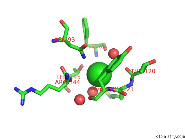
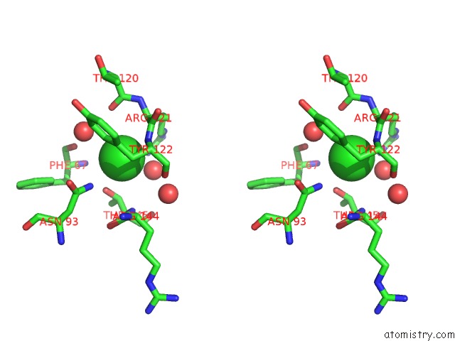
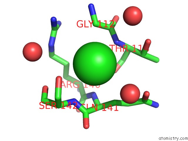
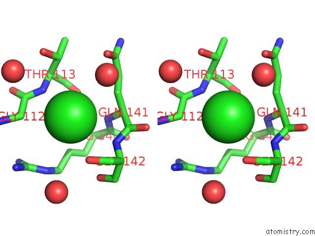
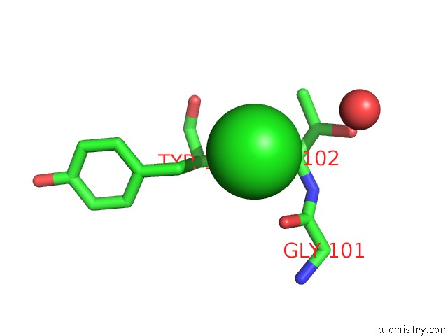
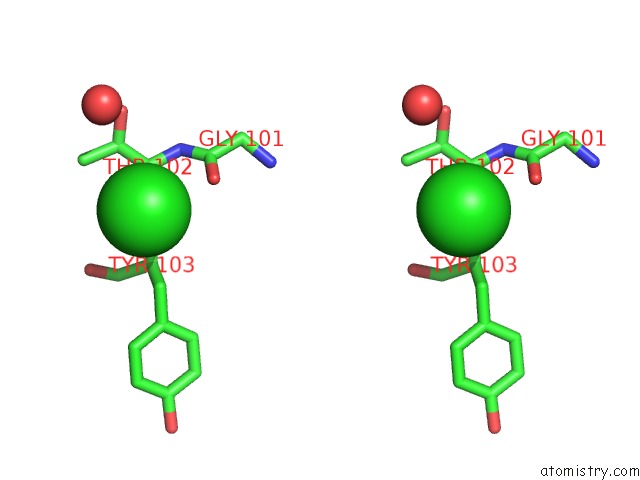
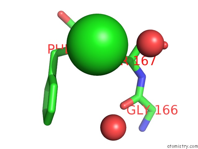
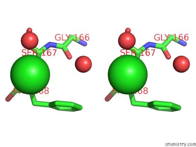
Chlorine binding site 1 out of 59 in 7dfn
Go back to
Chlorine binding site 1 out
of 59 in the Crystal Structure of Glycoside Hydrolase Family 11 Beta-Xylanase From Streptomyces Olivaceoviridis E-86 in Complex with Alpha-L- Arabinofuranosyl Xylotetraose
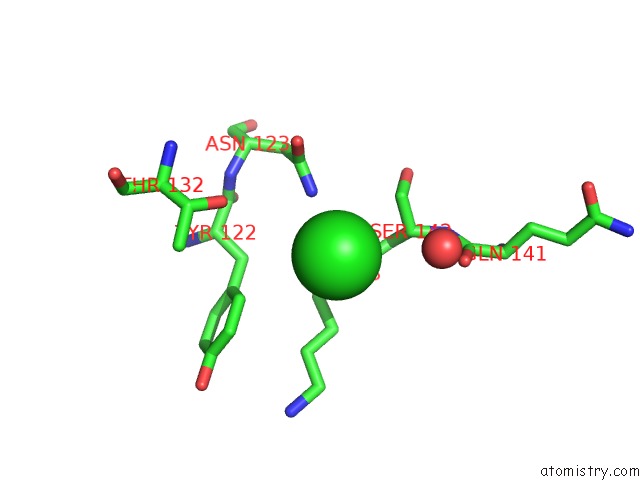
Mono view
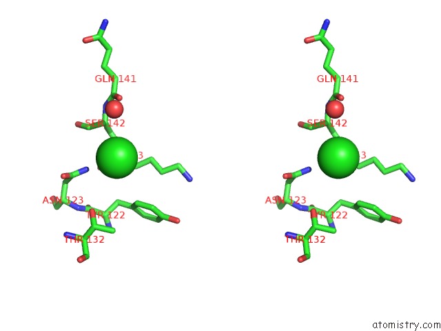
Stereo pair view
Mono view
Stereo pair view
A full contact list of Chlorine with other atoms in the Cl binding
site number 1 of Crystal Structure of Glycoside Hydrolase Family 11 Beta-Xylanase From Streptomyces Olivaceoviridis E-86 in Complex with Alpha-L- Arabinofuranosyl Xylotetraose within 5.0Å range:
|
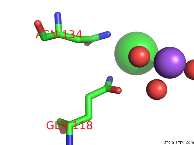
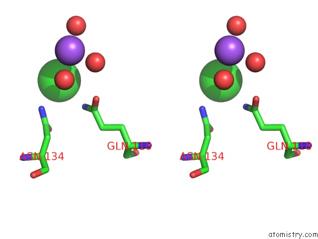
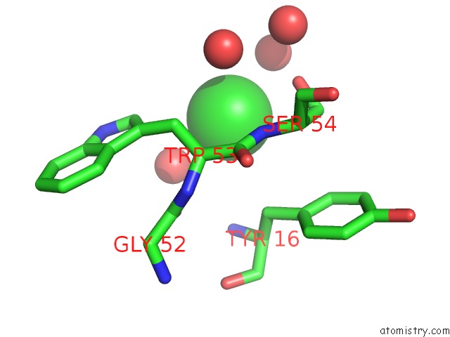
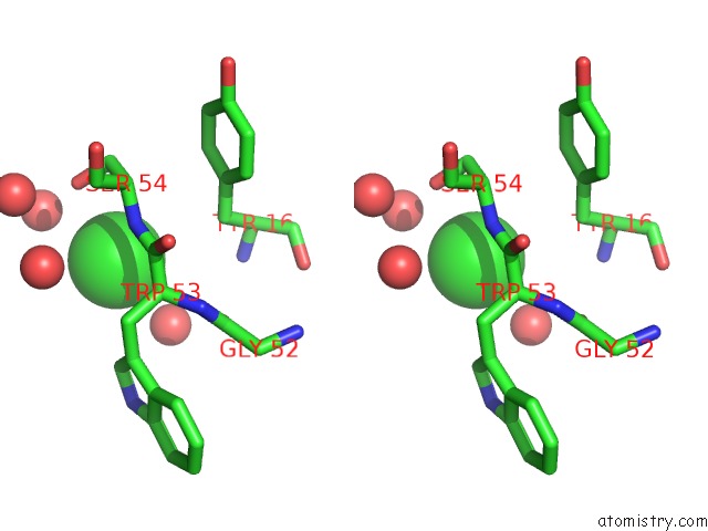
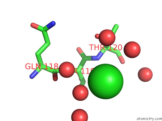
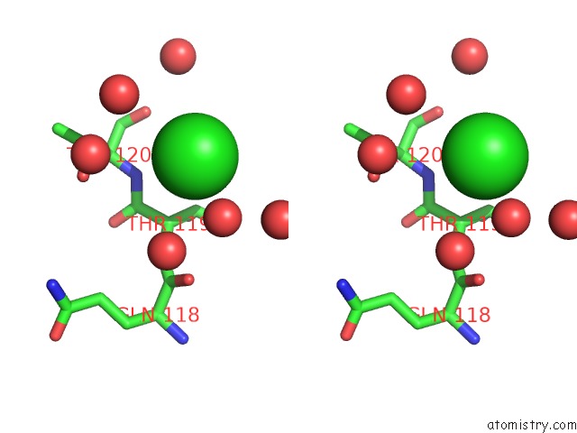
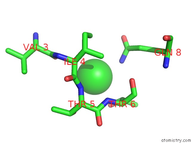
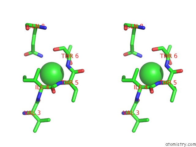
Chlorine binding site 2 out of 59 in 7dfn
Go back to
Chlorine binding site 2 out
of 59 in the Crystal Structure of Glycoside Hydrolase Family 11 Beta-Xylanase From Streptomyces Olivaceoviridis E-86 in Complex with Alpha-L- Arabinofuranosyl Xylotetraose
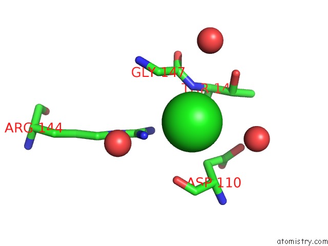
Mono view
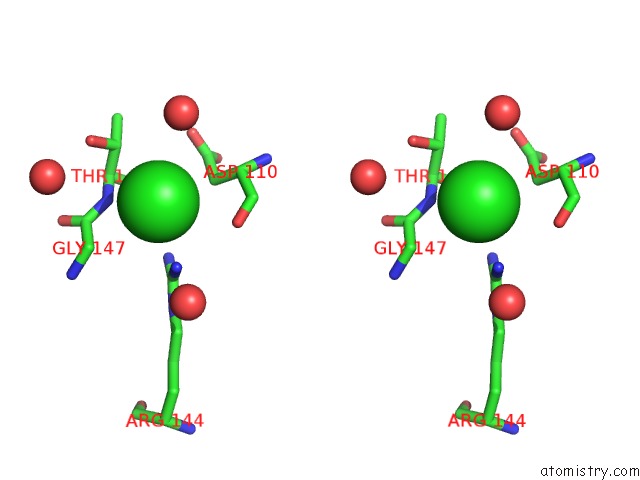
Stereo pair view
Mono view
Stereo pair view
A full contact list of Chlorine with other atoms in the Cl binding
site number 2 of Crystal Structure of Glycoside Hydrolase Family 11 Beta-Xylanase From Streptomyces Olivaceoviridis E-86 in Complex with Alpha-L- Arabinofuranosyl Xylotetraose within 5.0Å range:
|
Chlorine binding site 3 out of 59 in 7dfn
Go back to
Chlorine binding site 3 out
of 59 in the Crystal Structure of Glycoside Hydrolase Family 11 Beta-Xylanase From Streptomyces Olivaceoviridis E-86 in Complex with Alpha-L- Arabinofuranosyl Xylotetraose
Mono view
Stereo pair view
Mono view
Stereo pair view
A full contact list of Chlorine with other atoms in the Cl binding
site number 3 of Crystal Structure of Glycoside Hydrolase Family 11 Beta-Xylanase From Streptomyces Olivaceoviridis E-86 in Complex with Alpha-L- Arabinofuranosyl Xylotetraose within 5.0Å range:
|
Chlorine binding site 4 out of 59 in 7dfn
Go back to
Chlorine binding site 4 out
of 59 in the Crystal Structure of Glycoside Hydrolase Family 11 Beta-Xylanase From Streptomyces Olivaceoviridis E-86 in Complex with Alpha-L- Arabinofuranosyl Xylotetraose
Mono view
Stereo pair view
Mono view
Stereo pair view
A full contact list of Chlorine with other atoms in the Cl binding
site number 4 of Crystal Structure of Glycoside Hydrolase Family 11 Beta-Xylanase From Streptomyces Olivaceoviridis E-86 in Complex with Alpha-L- Arabinofuranosyl Xylotetraose within 5.0Å range:
|
Chlorine binding site 5 out of 59 in 7dfn
Go back to
Chlorine binding site 5 out
of 59 in the Crystal Structure of Glycoside Hydrolase Family 11 Beta-Xylanase From Streptomyces Olivaceoviridis E-86 in Complex with Alpha-L- Arabinofuranosyl Xylotetraose
Mono view
Stereo pair view
Mono view
Stereo pair view
A full contact list of Chlorine with other atoms in the Cl binding
site number 5 of Crystal Structure of Glycoside Hydrolase Family 11 Beta-Xylanase From Streptomyces Olivaceoviridis E-86 in Complex with Alpha-L- Arabinofuranosyl Xylotetraose within 5.0Å range:
|
Chlorine binding site 6 out of 59 in 7dfn
Go back to
Chlorine binding site 6 out
of 59 in the Crystal Structure of Glycoside Hydrolase Family 11 Beta-Xylanase From Streptomyces Olivaceoviridis E-86 in Complex with Alpha-L- Arabinofuranosyl Xylotetraose
Mono view
Stereo pair view
Mono view
Stereo pair view
A full contact list of Chlorine with other atoms in the Cl binding
site number 6 of Crystal Structure of Glycoside Hydrolase Family 11 Beta-Xylanase From Streptomyces Olivaceoviridis E-86 in Complex with Alpha-L- Arabinofuranosyl Xylotetraose within 5.0Å range:
|
Chlorine binding site 7 out of 59 in 7dfn
Go back to
Chlorine binding site 7 out
of 59 in the Crystal Structure of Glycoside Hydrolase Family 11 Beta-Xylanase From Streptomyces Olivaceoviridis E-86 in Complex with Alpha-L- Arabinofuranosyl Xylotetraose
Mono view
Stereo pair view
Mono view
Stereo pair view
A full contact list of Chlorine with other atoms in the Cl binding
site number 7 of Crystal Structure of Glycoside Hydrolase Family 11 Beta-Xylanase From Streptomyces Olivaceoviridis E-86 in Complex with Alpha-L- Arabinofuranosyl Xylotetraose within 5.0Å range:
|
Chlorine binding site 8 out of 59 in 7dfn
Go back to
Chlorine binding site 8 out
of 59 in the Crystal Structure of Glycoside Hydrolase Family 11 Beta-Xylanase From Streptomyces Olivaceoviridis E-86 in Complex with Alpha-L- Arabinofuranosyl Xylotetraose
Mono view
Stereo pair view
Mono view
Stereo pair view
A full contact list of Chlorine with other atoms in the Cl binding
site number 8 of Crystal Structure of Glycoside Hydrolase Family 11 Beta-Xylanase From Streptomyces Olivaceoviridis E-86 in Complex with Alpha-L- Arabinofuranosyl Xylotetraose within 5.0Å range:
|
Chlorine binding site 9 out of 59 in 7dfn
Go back to
Chlorine binding site 9 out
of 59 in the Crystal Structure of Glycoside Hydrolase Family 11 Beta-Xylanase From Streptomyces Olivaceoviridis E-86 in Complex with Alpha-L- Arabinofuranosyl Xylotetraose
Mono view
Stereo pair view
Mono view
Stereo pair view
A full contact list of Chlorine with other atoms in the Cl binding
site number 9 of Crystal Structure of Glycoside Hydrolase Family 11 Beta-Xylanase From Streptomyces Olivaceoviridis E-86 in Complex with Alpha-L- Arabinofuranosyl Xylotetraose within 5.0Å range:
|
Chlorine binding site 10 out of 59 in 7dfn
Go back to
Chlorine binding site 10 out
of 59 in the Crystal Structure of Glycoside Hydrolase Family 11 Beta-Xylanase From Streptomyces Olivaceoviridis E-86 in Complex with Alpha-L- Arabinofuranosyl Xylotetraose
Mono view
Stereo pair view
Mono view
Stereo pair view
A full contact list of Chlorine with other atoms in the Cl binding
site number 10 of Crystal Structure of Glycoside Hydrolase Family 11 Beta-Xylanase From Streptomyces Olivaceoviridis E-86 in Complex with Alpha-L- Arabinofuranosyl Xylotetraose within 5.0Å range:
|
Reference:
Z.Fujimoto,
K.Naomi,
K.Teramoto,
S.Tsutsui,
S.Kaneko.
Structure-Based Substrate Specificity of GH11 Xylanase From Streptomyces Olivaceoviridis E-86 To Be Published.
Page generated: Mon Jul 29 20:04:38 2024
Last articles
Zn in 9J0NZn in 9J0O
Zn in 9J0P
Zn in 9FJX
Zn in 9EKB
Zn in 9C0F
Zn in 9CAH
Zn in 9CH0
Zn in 9CH3
Zn in 9CH1