Chlorine »
PDB 7e4c-7epg »
7ea0 »
Chlorine in PDB 7ea0: Crystal Structure of Human Pyruvate Dehydrogenase Kinase 2 in Complex with Compound 1
Enzymatic activity of Crystal Structure of Human Pyruvate Dehydrogenase Kinase 2 in Complex with Compound 1
All present enzymatic activity of Crystal Structure of Human Pyruvate Dehydrogenase Kinase 2 in Complex with Compound 1:
2.7.11.2;
2.7.11.2;
Protein crystallography data
The structure of Crystal Structure of Human Pyruvate Dehydrogenase Kinase 2 in Complex with Compound 1, PDB code: 7ea0
was solved by
T.Orita,
S.Doi,
T.Iwanaga,
T.Adachi,
with X-Ray Crystallography technique. A brief refinement statistics is given in the table below:
| Resolution Low / High (Å) | 93.57 / 2.34 |
| Space group | P 64 |
| Cell size a, b, c (Å), α, β, γ (°) | 108.041, 108.041, 83.999, 90, 90, 120 |
| R / Rfree (%) | 19.3 / 21.5 |
Chlorine Binding Sites:
The binding sites of Chlorine atom in the Crystal Structure of Human Pyruvate Dehydrogenase Kinase 2 in Complex with Compound 1
(pdb code 7ea0). This binding sites where shown within
5.0 Angstroms radius around Chlorine atom.
In total only one binding site of Chlorine was determined in the Crystal Structure of Human Pyruvate Dehydrogenase Kinase 2 in Complex with Compound 1, PDB code: 7ea0:
In total only one binding site of Chlorine was determined in the Crystal Structure of Human Pyruvate Dehydrogenase Kinase 2 in Complex with Compound 1, PDB code: 7ea0:
Chlorine binding site 1 out of 1 in 7ea0
Go back to
Chlorine binding site 1 out
of 1 in the Crystal Structure of Human Pyruvate Dehydrogenase Kinase 2 in Complex with Compound 1
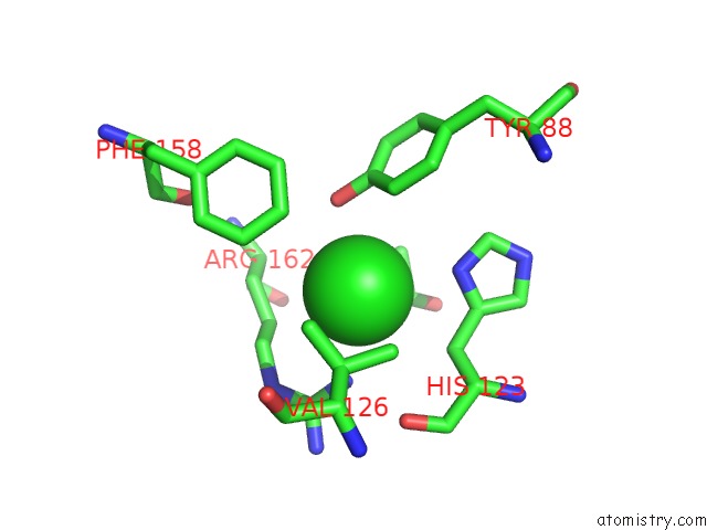
Mono view
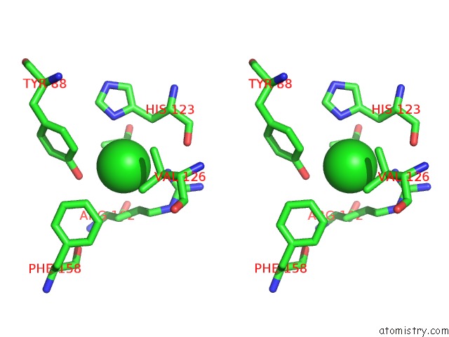
Stereo pair view
Mono view
Stereo pair view
A full contact list of Chlorine with other atoms in the Cl binding
site number 1 of Crystal Structure of Human Pyruvate Dehydrogenase Kinase 2 in Complex with Compound 1 within 5.0Å range:
|
Reference:
T.Akaki,
Y.Bessho,
T.Ito,
S.Fujioka,
M.Ubukata,
G.Mori,
K.Yamanaka,
T.Orita,
S.Doi,
T.Iwanaga,
K.Ikegashira,
Y.Hantani,
I.Nakanishi,
T.Adachi.
Fragment-Based Lead Discovery to Identify Novel Inhibitors That Target the Atp Binding Site of Pyruvate Dehydrogenase Kinases. Bioorg.Med.Chem. V. 44 16283 2021.
ISSN: ESSN 1464-3391
PubMed: 34274549
DOI: 10.1016/J.BMC.2021.116283
Page generated: Mon Jul 29 20:27:40 2024
ISSN: ESSN 1464-3391
PubMed: 34274549
DOI: 10.1016/J.BMC.2021.116283
Last articles
Zn in 9J0NZn in 9J0O
Zn in 9J0P
Zn in 9FJX
Zn in 9EKB
Zn in 9C0F
Zn in 9CAH
Zn in 9CH0
Zn in 9CH3
Zn in 9CH1