Chlorine »
PDB 7pe4-7ppr »
7pe4 »
Chlorine in PDB 7pe4: Crystal Structure of Mycobacterium Hassiacum Glucosyl-3- Phosphoglycerate Synthase at pH 5.5 in Complex with Udp-Glucose
Enzymatic activity of Crystal Structure of Mycobacterium Hassiacum Glucosyl-3- Phosphoglycerate Synthase at pH 5.5 in Complex with Udp-Glucose
All present enzymatic activity of Crystal Structure of Mycobacterium Hassiacum Glucosyl-3- Phosphoglycerate Synthase at pH 5.5 in Complex with Udp-Glucose:
2.4.1.266;
2.4.1.266;
Protein crystallography data
The structure of Crystal Structure of Mycobacterium Hassiacum Glucosyl-3- Phosphoglycerate Synthase at pH 5.5 in Complex with Udp-Glucose, PDB code: 7pe4
was solved by
A.Silva,
P.J.Babosa Pereira,
S.Macedo-Ribeiro,
with X-Ray Crystallography technique. A brief refinement statistics is given in the table below:
| Resolution Low / High (Å) | 46.58 / 2.05 |
| Space group | I 41 |
| Cell size a, b, c (Å), α, β, γ (°) | 101.887, 101.887, 122.098, 90, 90, 90 |
| R / Rfree (%) | 16 / 19.2 |
Other elements in 7pe4:
The structure of Crystal Structure of Mycobacterium Hassiacum Glucosyl-3- Phosphoglycerate Synthase at pH 5.5 in Complex with Udp-Glucose also contains other interesting chemical elements:
| Magnesium | (Mg) | 1 atom |
Chlorine Binding Sites:
The binding sites of Chlorine atom in the Crystal Structure of Mycobacterium Hassiacum Glucosyl-3- Phosphoglycerate Synthase at pH 5.5 in Complex with Udp-Glucose
(pdb code 7pe4). This binding sites where shown within
5.0 Angstroms radius around Chlorine atom.
In total 3 binding sites of Chlorine where determined in the Crystal Structure of Mycobacterium Hassiacum Glucosyl-3- Phosphoglycerate Synthase at pH 5.5 in Complex with Udp-Glucose, PDB code: 7pe4:
Jump to Chlorine binding site number: 1; 2; 3;
In total 3 binding sites of Chlorine where determined in the Crystal Structure of Mycobacterium Hassiacum Glucosyl-3- Phosphoglycerate Synthase at pH 5.5 in Complex with Udp-Glucose, PDB code: 7pe4:
Jump to Chlorine binding site number: 1; 2; 3;
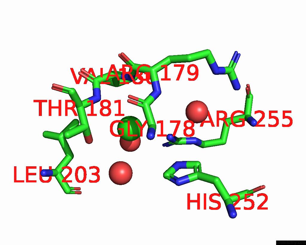
Chlorine binding site 1 out of 3 in 7pe4
Go back to
Chlorine binding site 1 out
of 3 in the Crystal Structure of Mycobacterium Hassiacum Glucosyl-3- Phosphoglycerate Synthase at pH 5.5 in Complex with Udp-Glucose
Mono view

Stereo pair view
Mono view
Stereo pair view
A full contact list of Chlorine with other atoms in the Cl binding
site number 1 of Crystal Structure of Mycobacterium Hassiacum Glucosyl-3- Phosphoglycerate Synthase at pH 5.5 in Complex with Udp-Glucose within 5.0Å range:
|
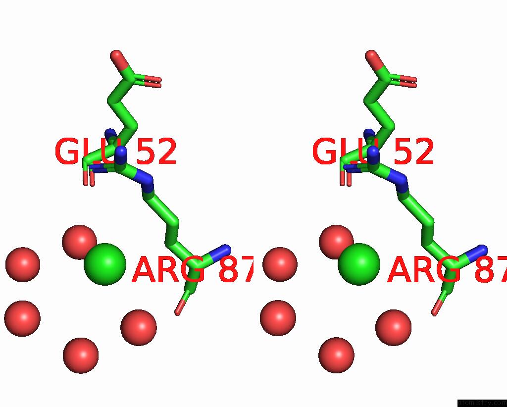
Chlorine binding site 2 out of 3 in 7pe4
Go back to
Chlorine binding site 2 out
of 3 in the Crystal Structure of Mycobacterium Hassiacum Glucosyl-3- Phosphoglycerate Synthase at pH 5.5 in Complex with Udp-Glucose

Mono view
Stereo pair view
Mono view
Stereo pair view
A full contact list of Chlorine with other atoms in the Cl binding
site number 2 of Crystal Structure of Mycobacterium Hassiacum Glucosyl-3- Phosphoglycerate Synthase at pH 5.5 in Complex with Udp-Glucose within 5.0Å range:
|
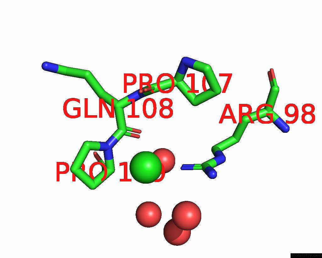
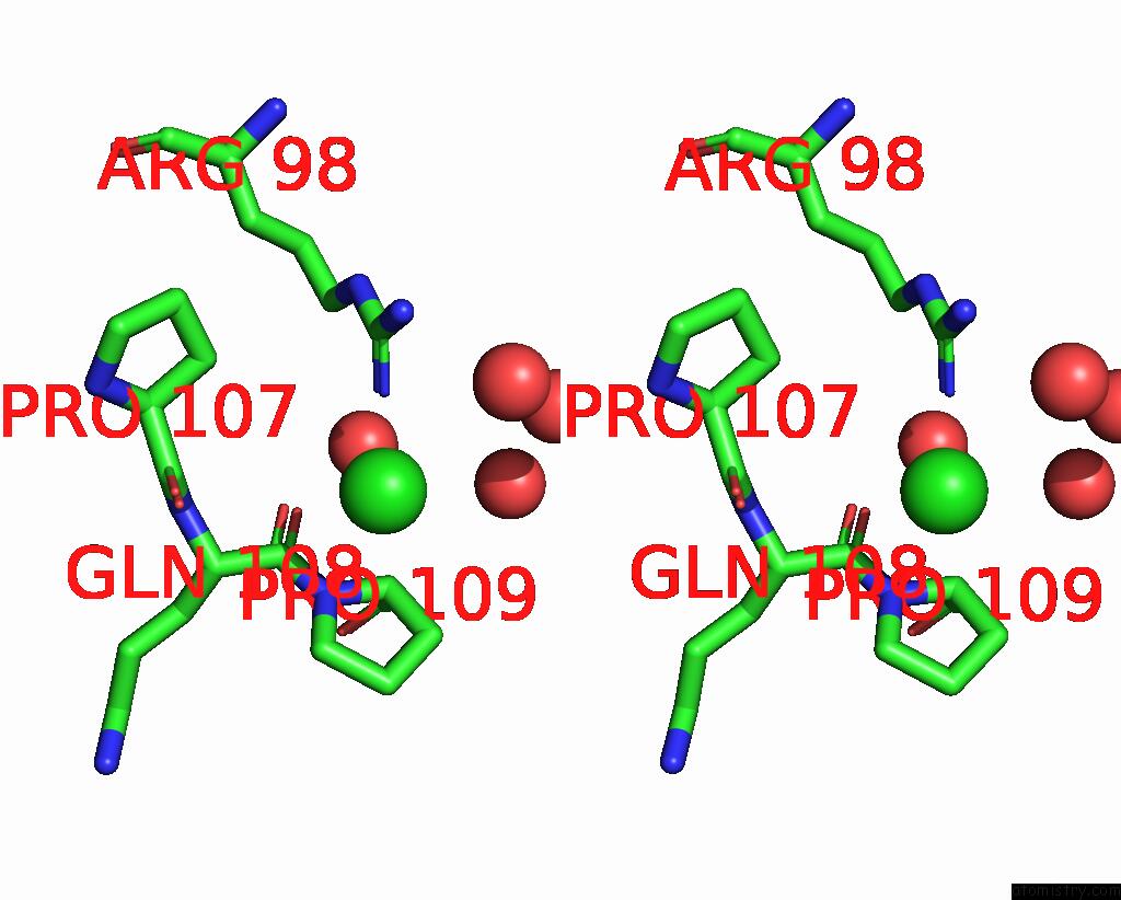
Chlorine binding site 3 out of 3 in 7pe4
Go back to
Chlorine binding site 3 out
of 3 in the Crystal Structure of Mycobacterium Hassiacum Glucosyl-3- Phosphoglycerate Synthase at pH 5.5 in Complex with Udp-Glucose
Mono view
Stereo pair view
Mono view
Stereo pair view
A full contact list of Chlorine with other atoms in the Cl binding
site number 3 of Crystal Structure of Mycobacterium Hassiacum Glucosyl-3- Phosphoglycerate Synthase at pH 5.5 in Complex with Udp-Glucose within 5.0Å range:
|
Reference:
A.Silva,
D.Nunes-Costa,
N.Empadinhas,
P.J.Babosa Pereira,
S.Macedo-Ribeiro.
Crystal Structure of Mycobacterium Hassiacum Glucosyl-3-Phosphoglycerate Synthase at pH 5.5 in Complex with Udp-Glucose To Be Published.
Page generated: Sun Jul 13 05:38:28 2025
Last articles
F in 4JJSF in 4JII
F in 4JDS
F in 4JAV
F in 4JA8
F in 4JBY
F in 4JB4
F in 4JAS
F in 4JA2
F in 4J3J