Chlorine »
PDB 7pps-7pw7 »
7ppv »
Chlorine in PDB 7ppv: Structure of Dife-Sulerythrin at 2.70 Mgy Total Absorbed Dose
Protein crystallography data
The structure of Structure of Dife-Sulerythrin at 2.70 Mgy Total Absorbed Dose, PDB code: 7ppv
was solved by
F.Lennartz,
M.S.Weiss,
with X-Ray Crystallography technique. A brief refinement statistics is given in the table below:
| Resolution Low / High (Å) | 38.55 / 1.36 |
| Space group | P 63 |
| Cell size a, b, c (Å), α, β, γ (°) | 72.15, 72.15, 97.98, 90, 90, 120 |
| R / Rfree (%) | 18.1 / 19.9 |
Other elements in 7ppv:
The structure of Structure of Dife-Sulerythrin at 2.70 Mgy Total Absorbed Dose also contains other interesting chemical elements:
| Iron | (Fe) | 6 atoms |
Chlorine Binding Sites:
The binding sites of Chlorine atom in the Structure of Dife-Sulerythrin at 2.70 Mgy Total Absorbed Dose
(pdb code 7ppv). This binding sites where shown within
5.0 Angstroms radius around Chlorine atom.
In total 2 binding sites of Chlorine where determined in the Structure of Dife-Sulerythrin at 2.70 Mgy Total Absorbed Dose, PDB code: 7ppv:
Jump to Chlorine binding site number: 1; 2;
In total 2 binding sites of Chlorine where determined in the Structure of Dife-Sulerythrin at 2.70 Mgy Total Absorbed Dose, PDB code: 7ppv:
Jump to Chlorine binding site number: 1; 2;
Chlorine binding site 1 out of 2 in 7ppv
Go back to
Chlorine binding site 1 out
of 2 in the Structure of Dife-Sulerythrin at 2.70 Mgy Total Absorbed Dose
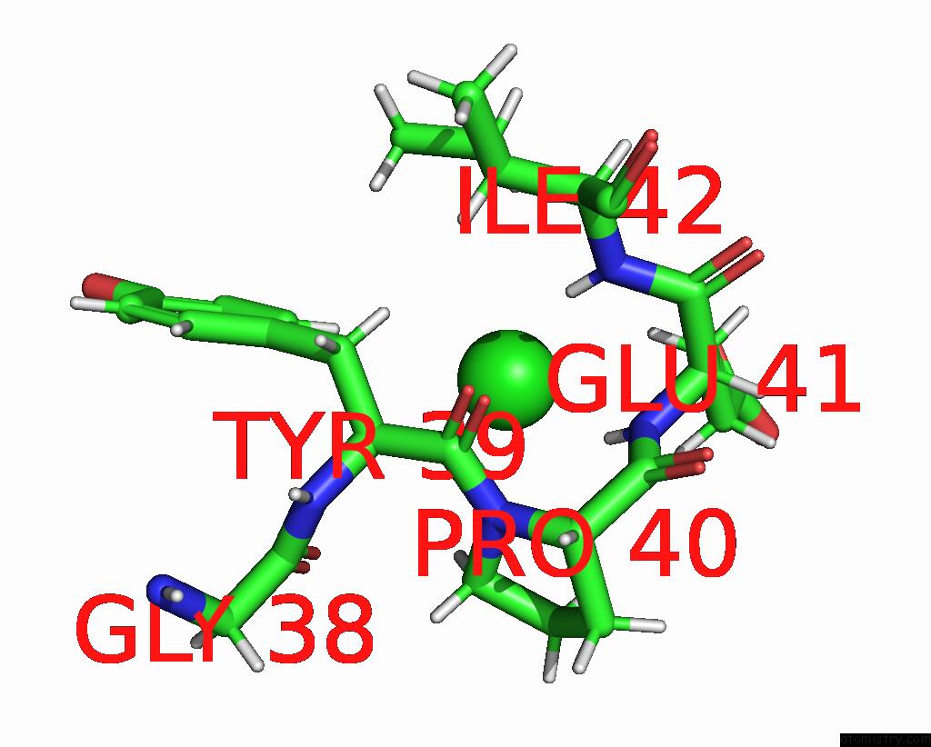
Mono view

Stereo pair view
Mono view
Stereo pair view
A full contact list of Chlorine with other atoms in the Cl binding
site number 1 of Structure of Dife-Sulerythrin at 2.70 Mgy Total Absorbed Dose within 5.0Å range:
|
Chlorine binding site 2 out of 2 in 7ppv
Go back to
Chlorine binding site 2 out
of 2 in the Structure of Dife-Sulerythrin at 2.70 Mgy Total Absorbed Dose
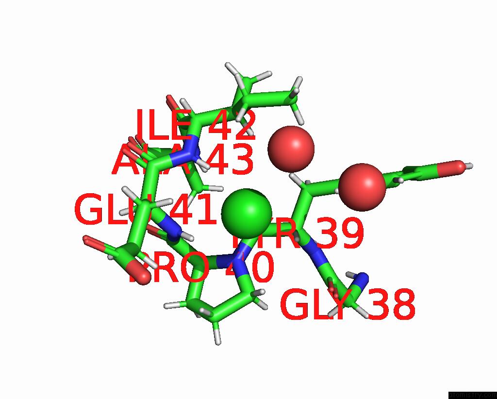
Mono view
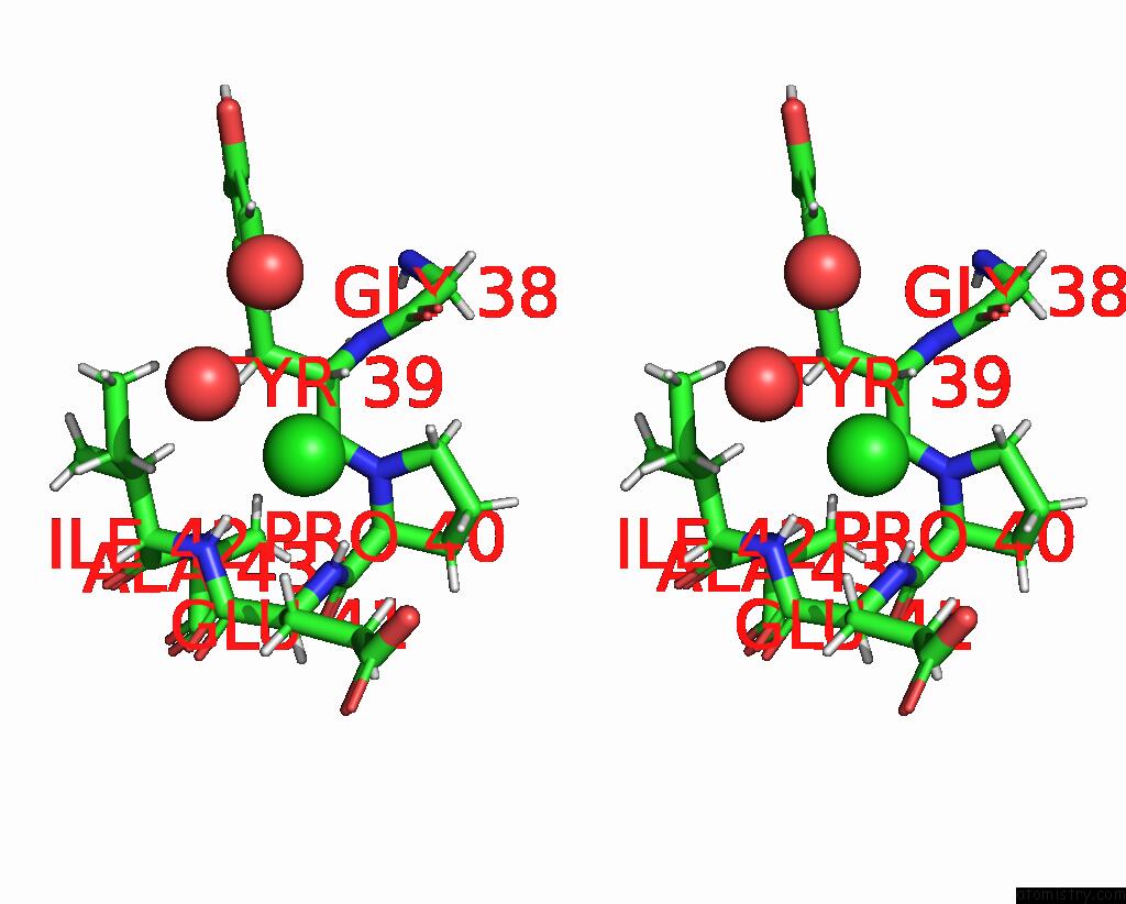
Stereo pair view
Mono view
Stereo pair view
A full contact list of Chlorine with other atoms in the Cl binding
site number 2 of Structure of Dife-Sulerythrin at 2.70 Mgy Total Absorbed Dose within 5.0Å range:
|
Reference:
F.Lennartz,
J.H.Jeoung,
S.Ruenger,
H.Dobbek,
M.S.Weiss.
Determining the Oxidation State of Elements By X-Ray Crystallography. Acta Crystallogr D Struct V. 78 238 2022BIOL.
ISSN: ISSN 2059-7983
PubMed: 35102889
DOI: 10.1107/S2059798321013048
Page generated: Sun Jul 13 05:49:44 2025
ISSN: ISSN 2059-7983
PubMed: 35102889
DOI: 10.1107/S2059798321013048
Last articles
F in 4LA6F in 4L9I
F in 4L3O
F in 4L7H
F in 4L8M
F in 4L7J
F in 4L4M
F in 4L6Q
F in 4L7F
F in 4L4L