Chlorine »
PDB 7pwd-7q1n »
7q0i »
Chlorine in PDB 7q0i: Crystal Structure of the N-Terminal Domain of Sars-Cov-2 Beta Variant Spike Glycoprotein in Complex with Beta-43
Protein crystallography data
The structure of Crystal Structure of the N-Terminal Domain of Sars-Cov-2 Beta Variant Spike Glycoprotein in Complex with Beta-43, PDB code: 7q0i
was solved by
D.Zhou,
J.Ren,
D.I.Stuart,
with X-Ray Crystallography technique. A brief refinement statistics is given in the table below:
| Resolution Low / High (Å) | 143.10 / 2.39 |
| Space group | P 1 21 1 |
| Cell size a, b, c (Å), α, β, γ (°) | 117.83, 66.964, 143.238, 90, 92.54, 90 |
| R / Rfree (%) | 20.2 / 24.2 |
Chlorine Binding Sites:
The binding sites of Chlorine atom in the Crystal Structure of the N-Terminal Domain of Sars-Cov-2 Beta Variant Spike Glycoprotein in Complex with Beta-43
(pdb code 7q0i). This binding sites where shown within
5.0 Angstroms radius around Chlorine atom.
In total 6 binding sites of Chlorine where determined in the Crystal Structure of the N-Terminal Domain of Sars-Cov-2 Beta Variant Spike Glycoprotein in Complex with Beta-43, PDB code: 7q0i:
Jump to Chlorine binding site number: 1; 2; 3; 4; 5; 6;
In total 6 binding sites of Chlorine where determined in the Crystal Structure of the N-Terminal Domain of Sars-Cov-2 Beta Variant Spike Glycoprotein in Complex with Beta-43, PDB code: 7q0i:
Jump to Chlorine binding site number: 1; 2; 3; 4; 5; 6;
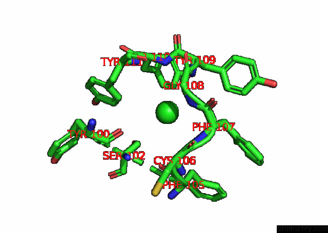
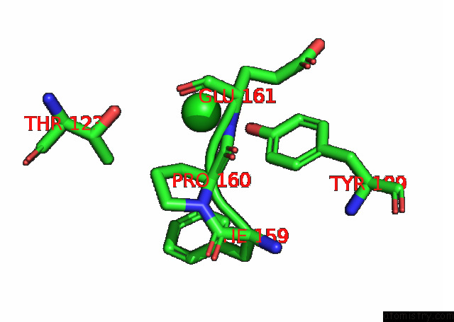
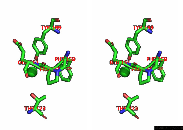
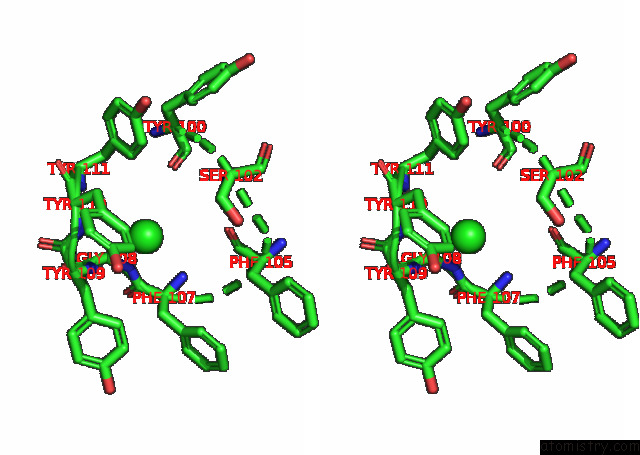
Chlorine binding site 1 out of 6 in 7q0i
Go back to
Chlorine binding site 1 out
of 6 in the Crystal Structure of the N-Terminal Domain of Sars-Cov-2 Beta Variant Spike Glycoprotein in Complex with Beta-43
Mono view
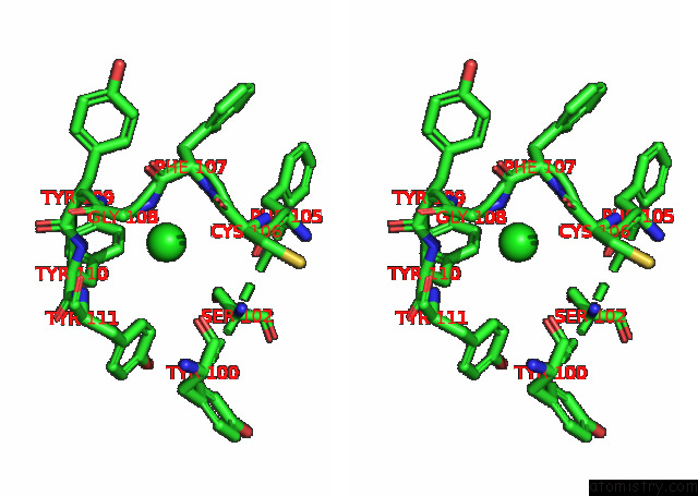
Stereo pair view
Mono view
Stereo pair view
A full contact list of Chlorine with other atoms in the Cl binding
site number 1 of Crystal Structure of the N-Terminal Domain of Sars-Cov-2 Beta Variant Spike Glycoprotein in Complex with Beta-43 within 5.0Å range:
|
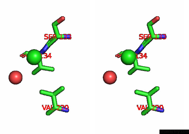
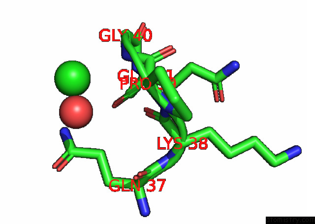
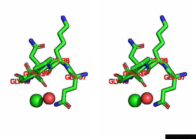
Chlorine binding site 2 out of 6 in 7q0i
Go back to
Chlorine binding site 2 out
of 6 in the Crystal Structure of the N-Terminal Domain of Sars-Cov-2 Beta Variant Spike Glycoprotein in Complex with Beta-43
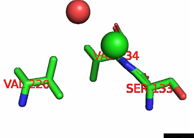
Mono view
Stereo pair view
Mono view
Stereo pair view
A full contact list of Chlorine with other atoms in the Cl binding
site number 2 of Crystal Structure of the N-Terminal Domain of Sars-Cov-2 Beta Variant Spike Glycoprotein in Complex with Beta-43 within 5.0Å range:
|
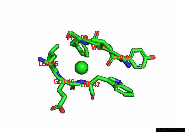
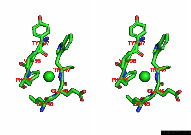
Chlorine binding site 3 out of 6 in 7q0i
Go back to
Chlorine binding site 3 out
of 6 in the Crystal Structure of the N-Terminal Domain of Sars-Cov-2 Beta Variant Spike Glycoprotein in Complex with Beta-43
Mono view
Stereo pair view
Mono view
Stereo pair view
A full contact list of Chlorine with other atoms in the Cl binding
site number 3 of Crystal Structure of the N-Terminal Domain of Sars-Cov-2 Beta Variant Spike Glycoprotein in Complex with Beta-43 within 5.0Å range:
|
Chlorine binding site 4 out of 6 in 7q0i
Go back to
Chlorine binding site 4 out
of 6 in the Crystal Structure of the N-Terminal Domain of Sars-Cov-2 Beta Variant Spike Glycoprotein in Complex with Beta-43
Mono view
Stereo pair view
Mono view
Stereo pair view
A full contact list of Chlorine with other atoms in the Cl binding
site number 4 of Crystal Structure of the N-Terminal Domain of Sars-Cov-2 Beta Variant Spike Glycoprotein in Complex with Beta-43 within 5.0Å range:
|
Chlorine binding site 5 out of 6 in 7q0i
Go back to
Chlorine binding site 5 out
of 6 in the Crystal Structure of the N-Terminal Domain of Sars-Cov-2 Beta Variant Spike Glycoprotein in Complex with Beta-43

Mono view
Stereo pair view
Mono view
Stereo pair view
A full contact list of Chlorine with other atoms in the Cl binding
site number 5 of Crystal Structure of the N-Terminal Domain of Sars-Cov-2 Beta Variant Spike Glycoprotein in Complex with Beta-43 within 5.0Å range:
|
Chlorine binding site 6 out of 6 in 7q0i
Go back to
Chlorine binding site 6 out
of 6 in the Crystal Structure of the N-Terminal Domain of Sars-Cov-2 Beta Variant Spike Glycoprotein in Complex with Beta-43
Mono view
Stereo pair view
Mono view
Stereo pair view
A full contact list of Chlorine with other atoms in the Cl binding
site number 6 of Crystal Structure of the N-Terminal Domain of Sars-Cov-2 Beta Variant Spike Glycoprotein in Complex with Beta-43 within 5.0Å range:
|
Reference:
C.Liu,
D.Zhou,
R.Nutalai,
H.M.Duyvesteyn,
A.Tuekprakhon,
H.M.Ginn,
W.Dejnirattisai,
P.Supasa,
A.J.Mentzer,
B.Wang,
J.B.Case,
Y.Zhao,
D.T.Skelly,
R.E.Chen,
S.A.Johnson,
T.G.Ritter,
C.Mason,
T.Malik,
N.Temperton,
N.G.Paterson,
M.A.Williams,
D.R.Hall,
D.K.Clare,
A.Howe,
P.J.Goulder,
E.E.Fry,
M.S.Diamond,
J.Mongkolsapaya,
J.Ren,
D.I.Stuart,
G.R.Screaton.
The Antibody Response to Sars-Cov-2 Beta Underscores the Antigenic Distance to Other Variants Cell Host Microbe 2021.
ISSN: ESSN 1934-6069
DOI: 10.1016/J.CHOM.2021.11.013
Page generated: Sun Jul 13 06:04:16 2025
ISSN: ESSN 1934-6069
DOI: 10.1016/J.CHOM.2021.11.013
Last articles
Fe in 2YXOFe in 2YRS
Fe in 2YXC
Fe in 2YNM
Fe in 2YVJ
Fe in 2YP1
Fe in 2YU2
Fe in 2YU1
Fe in 2YQB
Fe in 2YOO