Chlorine »
PDB 7qou-7r44 »
7r3r »
Chlorine in PDB 7r3r: Crystal Structure of Ctx-M-15 G238C Mutant Apoenzyme
Enzymatic activity of Crystal Structure of Ctx-M-15 G238C Mutant Apoenzyme
All present enzymatic activity of Crystal Structure of Ctx-M-15 G238C Mutant Apoenzyme:
3.5.2.6;
3.5.2.6;
Protein crystallography data
The structure of Crystal Structure of Ctx-M-15 G238C Mutant Apoenzyme, PDB code: 7r3r
was solved by
C.L.Tooke,
P.Hinchliffe,
J.Spencer,
with X-Ray Crystallography technique. A brief refinement statistics is given in the table below:
| Resolution Low / High (Å) | 58.94 / 1.17 |
| Space group | P 21 21 21 |
| Cell size a, b, c (Å), α, β, γ (°) | 44.553, 45.653, 117.876, 90, 90, 90 |
| R / Rfree (%) | 15.4 / 18.7 |
Chlorine Binding Sites:
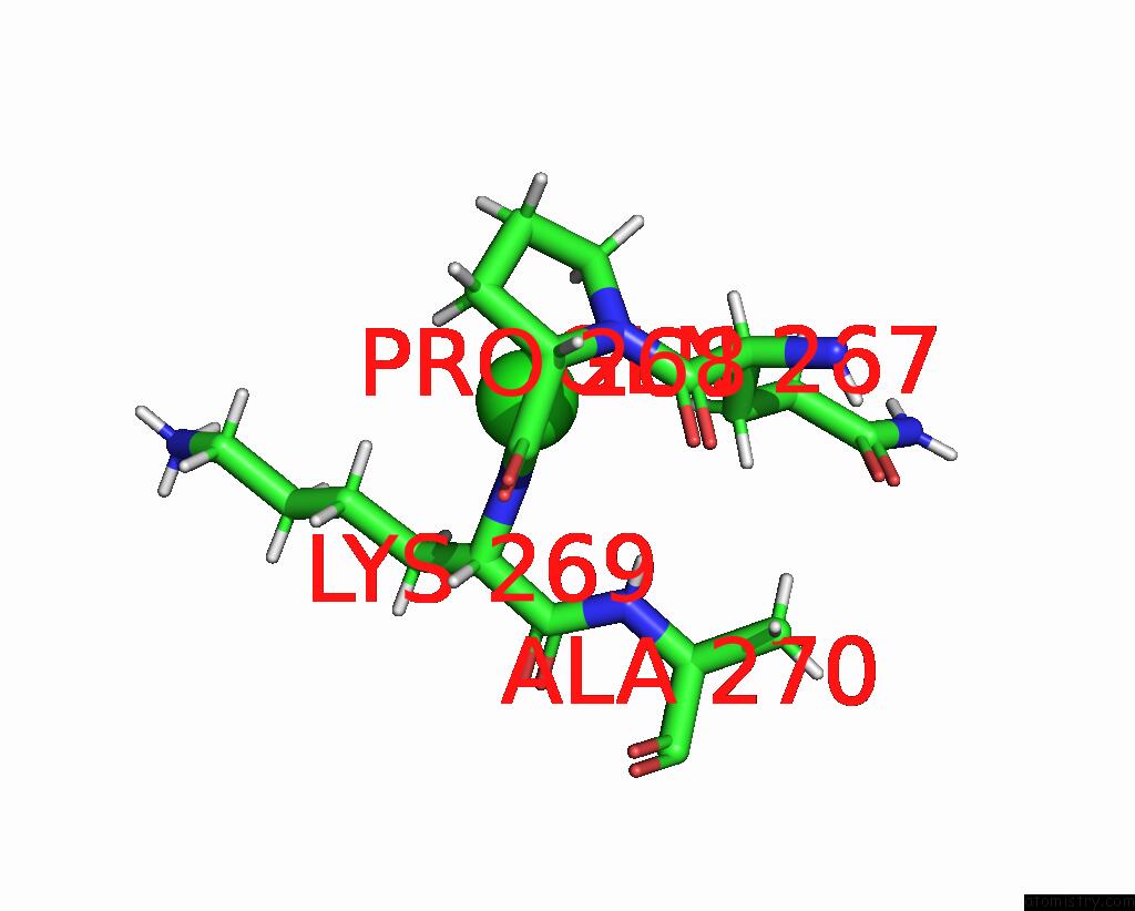
The binding sites of Chlorine atom in the Crystal Structure of Ctx-M-15 G238C Mutant Apoenzyme
(pdb code 7r3r). This binding sites where shown within
5.0 Angstroms radius around Chlorine atom.
In total only one binding site of Chlorine was determined in the Crystal Structure of Ctx-M-15 G238C Mutant Apoenzyme, PDB code: 7r3r:
In total only one binding site of Chlorine was determined in the Crystal Structure of Ctx-M-15 G238C Mutant Apoenzyme, PDB code: 7r3r:
Chlorine binding site 1 out of 1 in 7r3r
Go back to
Chlorine binding site 1 out
of 1 in the Crystal Structure of Ctx-M-15 G238C Mutant Apoenzyme
Mono view

Stereo pair view
Mono view
Stereo pair view
A full contact list of Chlorine with other atoms in the Cl binding
site number 1 of Crystal Structure of Ctx-M-15 G238C Mutant Apoenzyme within 5.0Å range:
|
Reference:
P.Hinchliffe,
C.L.Tooke,
C.R.Bethel,
B.Wang,
C.Arthur,
K.J.Heesom,
S.Shapiro,
D.M.Schlatzer,
K.M.Papp-Wallace,
R.A.Bonomo,
J.Spencer.
Penicillanic Acid Sulfones Inactivate the Extended-Spectrum Beta-Lactamase Ctx-M-15 Through Formation of A Serine-Lysine Cross-Link: An Alternative Mechanism of Beta-Lactamase Inhibition. Mbio V. 13 79321 2022.
ISSN: ESSN 2150-7511
PubMed: 35612361
DOI: 10.1128/MBIO.01793-21
Page generated: Tue Jul 30 03:28:44 2024
ISSN: ESSN 2150-7511
PubMed: 35612361
DOI: 10.1128/MBIO.01793-21
Last articles
Zn in 9JYWZn in 9IR4
Zn in 9IR3
Zn in 9GMX
Zn in 9GMW
Zn in 9JEJ
Zn in 9ERF
Zn in 9ERE
Zn in 9EGV
Zn in 9EGW