Chlorine »
PDB 7qou-7r44 »
7r3t »
Chlorine in PDB 7r3t: Crystal Structure of the Dimeric C-Terminal BIG_2-CBM56 Domains From Paenibacillus Illinoisensis (Bacillus Circulans IAM1165) Beta-1,3- Glucanase H
Protein crystallography data
The structure of Crystal Structure of the Dimeric C-Terminal BIG_2-CBM56 Domains From Paenibacillus Illinoisensis (Bacillus Circulans IAM1165) Beta-1,3- Glucanase H, PDB code: 7r3t
was solved by
S.Najmudin,
I.Venditto,
C.M.G.A.Fontes,
P.Bule,
with X-Ray Crystallography technique. A brief refinement statistics is given in the table below:
| Resolution Low / High (Å) | 45.80 / 2.11 |
| Space group | P 2 21 21 |
| Cell size a, b, c (Å), α, β, γ (°) | 50.63, 76.38, 106.87, 90, 90, 90 |
| R / Rfree (%) | 22.1 / 29.6 |
Chlorine Binding Sites:
The binding sites of Chlorine atom in the Crystal Structure of the Dimeric C-Terminal BIG_2-CBM56 Domains From Paenibacillus Illinoisensis (Bacillus Circulans IAM1165) Beta-1,3- Glucanase H
(pdb code 7r3t). This binding sites where shown within
5.0 Angstroms radius around Chlorine atom.
In total 3 binding sites of Chlorine where determined in the Crystal Structure of the Dimeric C-Terminal BIG_2-CBM56 Domains From Paenibacillus Illinoisensis (Bacillus Circulans IAM1165) Beta-1,3- Glucanase H, PDB code: 7r3t:
Jump to Chlorine binding site number: 1; 2; 3;
In total 3 binding sites of Chlorine where determined in the Crystal Structure of the Dimeric C-Terminal BIG_2-CBM56 Domains From Paenibacillus Illinoisensis (Bacillus Circulans IAM1165) Beta-1,3- Glucanase H, PDB code: 7r3t:
Jump to Chlorine binding site number: 1; 2; 3;
Chlorine binding site 1 out of 3 in 7r3t
Go back to
Chlorine binding site 1 out
of 3 in the Crystal Structure of the Dimeric C-Terminal BIG_2-CBM56 Domains From Paenibacillus Illinoisensis (Bacillus Circulans IAM1165) Beta-1,3- Glucanase H
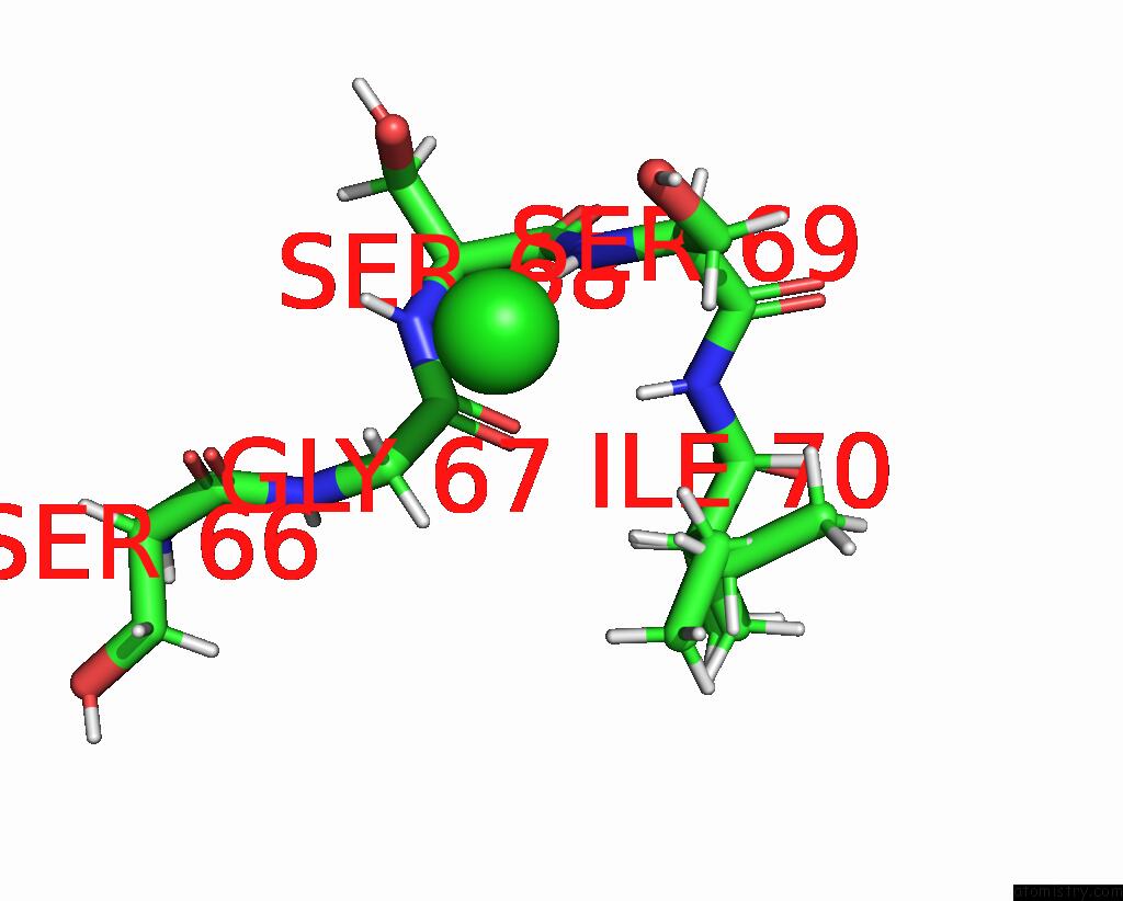
Mono view

Stereo pair view
Mono view
Stereo pair view
A full contact list of Chlorine with other atoms in the Cl binding
site number 1 of Crystal Structure of the Dimeric C-Terminal BIG_2-CBM56 Domains From Paenibacillus Illinoisensis (Bacillus Circulans IAM1165) Beta-1,3- Glucanase H within 5.0Å range:
|
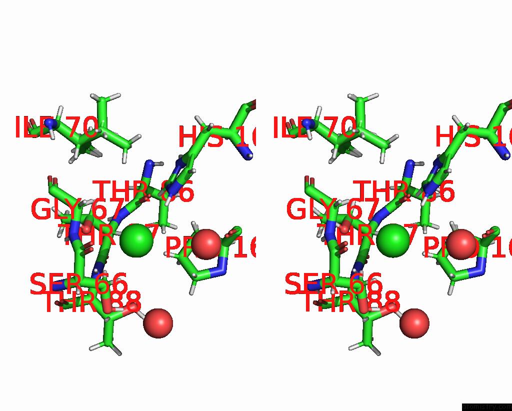
Chlorine binding site 2 out of 3 in 7r3t
Go back to
Chlorine binding site 2 out
of 3 in the Crystal Structure of the Dimeric C-Terminal BIG_2-CBM56 Domains From Paenibacillus Illinoisensis (Bacillus Circulans IAM1165) Beta-1,3- Glucanase H
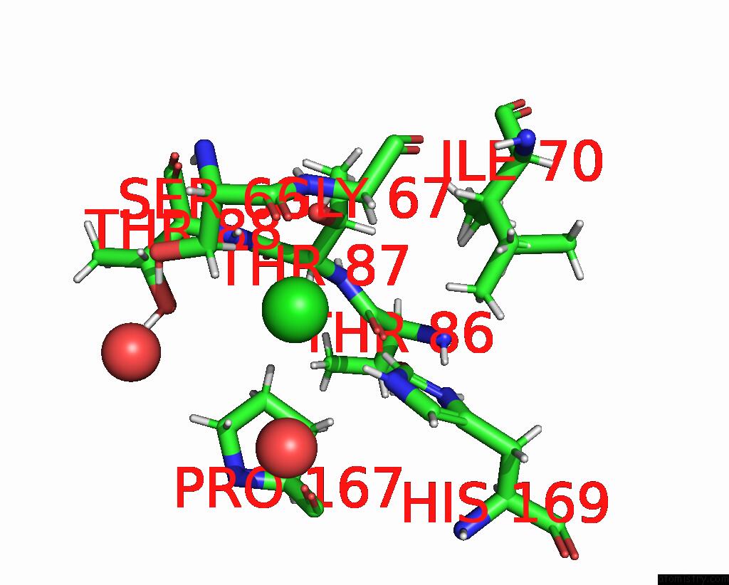
Mono view
Stereo pair view
Mono view
Stereo pair view
A full contact list of Chlorine with other atoms in the Cl binding
site number 2 of Crystal Structure of the Dimeric C-Terminal BIG_2-CBM56 Domains From Paenibacillus Illinoisensis (Bacillus Circulans IAM1165) Beta-1,3- Glucanase H within 5.0Å range:
|
Chlorine binding site 3 out of 3 in 7r3t
Go back to
Chlorine binding site 3 out
of 3 in the Crystal Structure of the Dimeric C-Terminal BIG_2-CBM56 Domains From Paenibacillus Illinoisensis (Bacillus Circulans IAM1165) Beta-1,3- Glucanase H
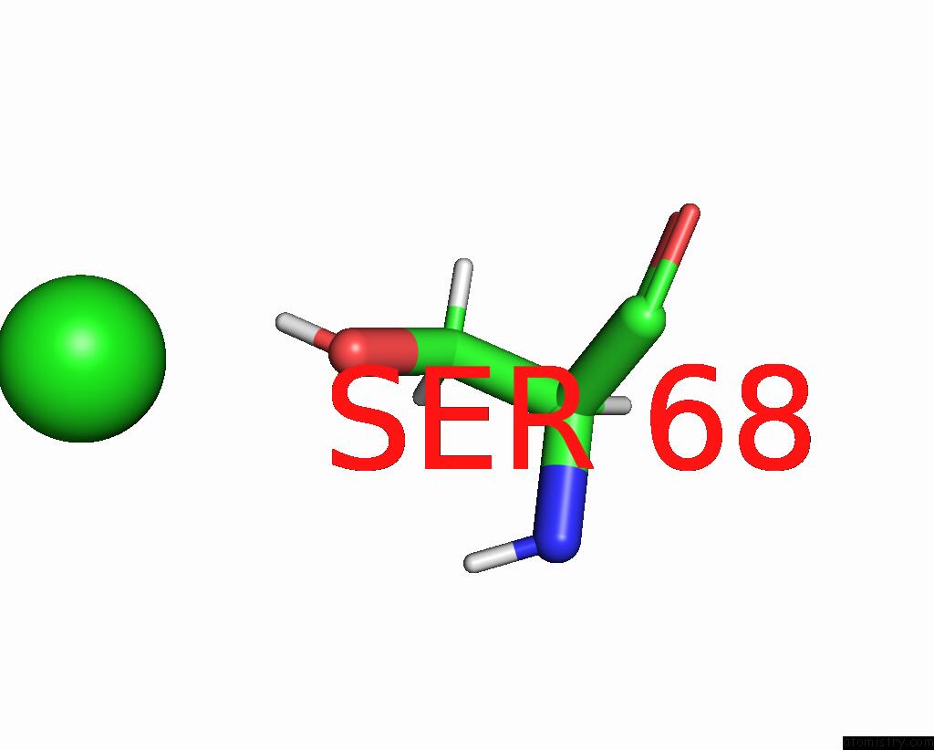
Mono view

Stereo pair view
Mono view
Stereo pair view
A full contact list of Chlorine with other atoms in the Cl binding
site number 3 of Crystal Structure of the Dimeric C-Terminal BIG_2-CBM56 Domains From Paenibacillus Illinoisensis (Bacillus Circulans IAM1165) Beta-1,3- Glucanase H within 5.0Å range:
|
Reference:
S.Najmudin,
I.Venditto,
V.R.Pires,
C.Caseiro,
M.A.S.Correia,
M.J.Romao,
A.L.Carvalho,
C.M.G.A.Fontes,
P.Bule.
Structural and Biochemical Characterization of C-Terminal BIG_2-CBM56 Domains of Paenibacillus Illinoisensis IAM1165 Beta-1,3-Glucanase H and Paenibacillus Sp CBM56 To Be Published.
Page generated: Tue Jul 30 03:28:44 2024
Last articles
Zn in 9JYWZn in 9IR4
Zn in 9IR3
Zn in 9GMX
Zn in 9GMW
Zn in 9JEJ
Zn in 9ERF
Zn in 9ERE
Zn in 9EGV
Zn in 9EGW