Chlorine »
PDB 7uh9-7uvn »
7uul »
Chlorine in PDB 7uul: Crystal Structure of Aminoglycoside Resistance Enzyme Apma, Complex with Kanamycin B and Coenzyme A
Protein crystallography data
The structure of Crystal Structure of Aminoglycoside Resistance Enzyme Apma, Complex with Kanamycin B and Coenzyme A, PDB code: 7uul
was solved by
P.J.Stogios,
E.Evdokimova,
R.Di Leo,
E.Bordeleau,
G.D.Wright,
A.Savchenko,
A.Joachimiak,
K.J.F.Satchell,
Center For Structural Genomics Ofinfectious Diseases (Csgid),
with X-Ray Crystallography technique. A brief refinement statistics is given in the table below:
| Resolution Low / High (Å) | 29.47 / 2.26 |
| Space group | P 21 21 21 |
| Cell size a, b, c (Å), α, β, γ (°) | 101.093, 131.685, 157.915, 90, 90, 90 |
| R / Rfree (%) | 17.2 / 23.2 |
Chlorine Binding Sites:
The binding sites of Chlorine atom in the Crystal Structure of Aminoglycoside Resistance Enzyme Apma, Complex with Kanamycin B and Coenzyme A
(pdb code 7uul). This binding sites where shown within
5.0 Angstroms radius around Chlorine atom.
In total only one binding site of Chlorine was determined in the Crystal Structure of Aminoglycoside Resistance Enzyme Apma, Complex with Kanamycin B and Coenzyme A, PDB code: 7uul:
In total only one binding site of Chlorine was determined in the Crystal Structure of Aminoglycoside Resistance Enzyme Apma, Complex with Kanamycin B and Coenzyme A, PDB code: 7uul:
Chlorine binding site 1 out of 1 in 7uul
Go back to
Chlorine binding site 1 out
of 1 in the Crystal Structure of Aminoglycoside Resistance Enzyme Apma, Complex with Kanamycin B and Coenzyme A
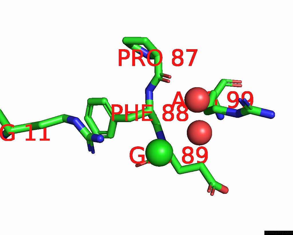
Mono view
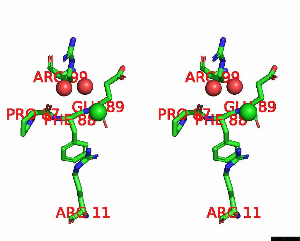
Stereo pair view
Mono view
Stereo pair view
A full contact list of Chlorine with other atoms in the Cl binding
site number 1 of Crystal Structure of Aminoglycoside Resistance Enzyme Apma, Complex with Kanamycin B and Coenzyme A within 5.0Å range:
|
Reference:
P.J.Stogios,
P.J.Stogios,
E.Evdokimova,
R.Di Leo,
E.Bordeleau,
G.D.Wright,
A.Savchenko,
A.Joachimiak,
K.J.F.Satchell,
Center For Structural Genomics Ofinfectious Diseases (Csgid).
N/A N/A.
Page generated: Tue Jul 30 05:11:38 2024
Last articles
Zn in 9JYWZn in 9IR4
Zn in 9IR3
Zn in 9GMX
Zn in 9GMW
Zn in 9JEJ
Zn in 9ERF
Zn in 9ERE
Zn in 9EGV
Zn in 9EGW