Chlorine »
PDB 7xwr-7yq2 »
7ynv »
Chlorine in PDB 7ynv: Crystal Structure of Photolysed Hen Egg White Lysozyme Introduced with O-(2-Nitrobenzyl)-L-Tyrosine
Enzymatic activity of Crystal Structure of Photolysed Hen Egg White Lysozyme Introduced with O-(2-Nitrobenzyl)-L-Tyrosine
All present enzymatic activity of Crystal Structure of Photolysed Hen Egg White Lysozyme Introduced with O-(2-Nitrobenzyl)-L-Tyrosine:
3.2.1.17;
3.2.1.17;
Protein crystallography data
The structure of Crystal Structure of Photolysed Hen Egg White Lysozyme Introduced with O-(2-Nitrobenzyl)-L-Tyrosine, PDB code: 7ynv
was solved by
T.Hosaka,
M.Shirouzu,
with X-Ray Crystallography technique. A brief refinement statistics is given in the table below:
| Resolution Low / High (Å) | 39.14 / 1.39 |
| Space group | P 43 21 2 |
| Cell size a, b, c (Å), α, β, γ (°) | 78.28, 78.28, 37.37, 90, 90, 90 |
| R / Rfree (%) | 17.4 / 19.2 |
Other elements in 7ynv:
The structure of Crystal Structure of Photolysed Hen Egg White Lysozyme Introduced with O-(2-Nitrobenzyl)-L-Tyrosine also contains other interesting chemical elements:
| Sodium | (Na) | 1 atom |
Chlorine Binding Sites:
The binding sites of Chlorine atom in the Crystal Structure of Photolysed Hen Egg White Lysozyme Introduced with O-(2-Nitrobenzyl)-L-Tyrosine
(pdb code 7ynv). This binding sites where shown within
5.0 Angstroms radius around Chlorine atom.
In total 5 binding sites of Chlorine where determined in the Crystal Structure of Photolysed Hen Egg White Lysozyme Introduced with O-(2-Nitrobenzyl)-L-Tyrosine, PDB code: 7ynv:
Jump to Chlorine binding site number: 1; 2; 3; 4; 5;
In total 5 binding sites of Chlorine where determined in the Crystal Structure of Photolysed Hen Egg White Lysozyme Introduced with O-(2-Nitrobenzyl)-L-Tyrosine, PDB code: 7ynv:
Jump to Chlorine binding site number: 1; 2; 3; 4; 5;
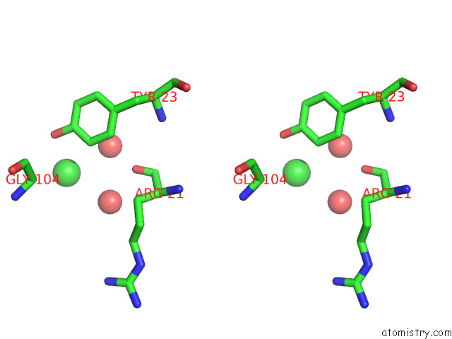
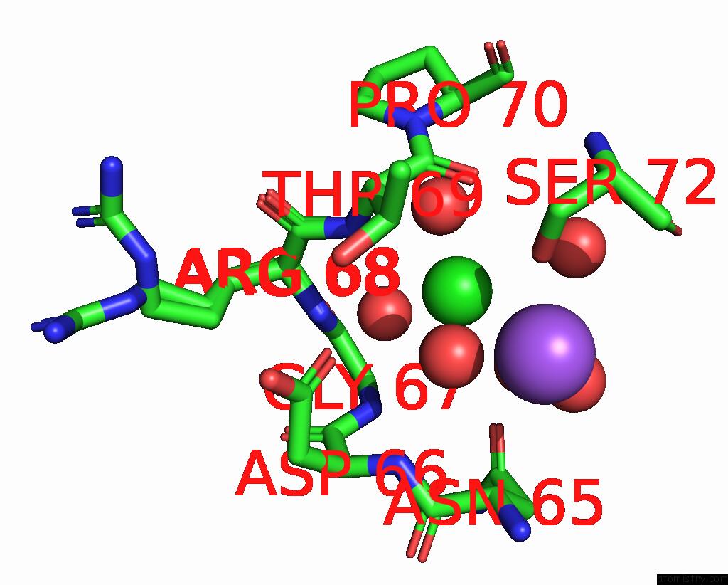
Chlorine binding site 1 out of 5 in 7ynv
Go back to
Chlorine binding site 1 out
of 5 in the Crystal Structure of Photolysed Hen Egg White Lysozyme Introduced with O-(2-Nitrobenzyl)-L-Tyrosine

Mono view
Stereo pair view
Mono view
Stereo pair view
A full contact list of Chlorine with other atoms in the Cl binding
site number 1 of Crystal Structure of Photolysed Hen Egg White Lysozyme Introduced with O-(2-Nitrobenzyl)-L-Tyrosine within 5.0Å range:
|
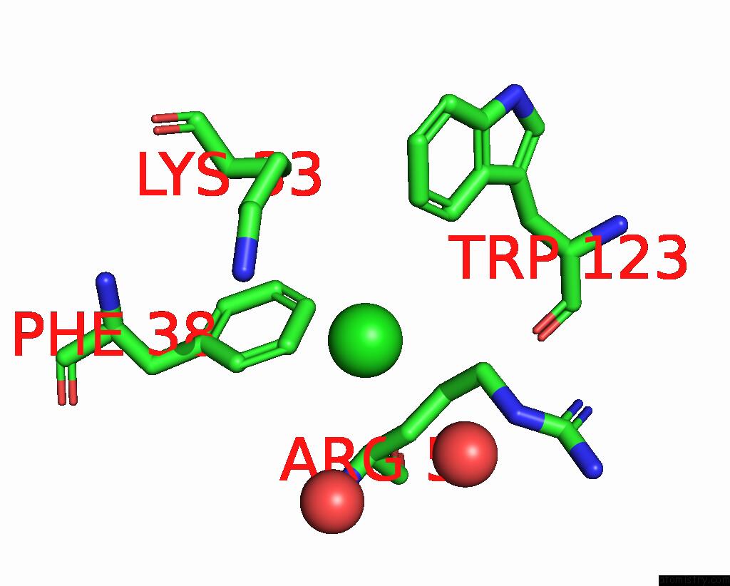
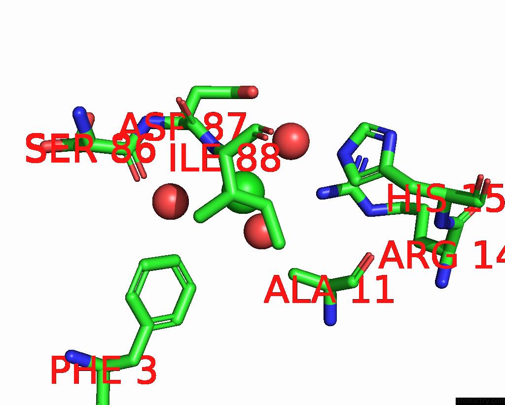
Chlorine binding site 2 out of 5 in 7ynv
Go back to
Chlorine binding site 2 out
of 5 in the Crystal Structure of Photolysed Hen Egg White Lysozyme Introduced with O-(2-Nitrobenzyl)-L-Tyrosine
Mono view

Stereo pair view
Mono view
Stereo pair view
A full contact list of Chlorine with other atoms in the Cl binding
site number 2 of Crystal Structure of Photolysed Hen Egg White Lysozyme Introduced with O-(2-Nitrobenzyl)-L-Tyrosine within 5.0Å range:
|
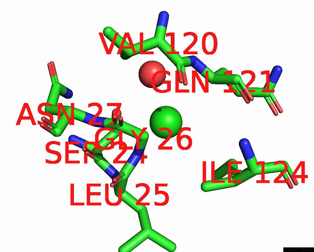
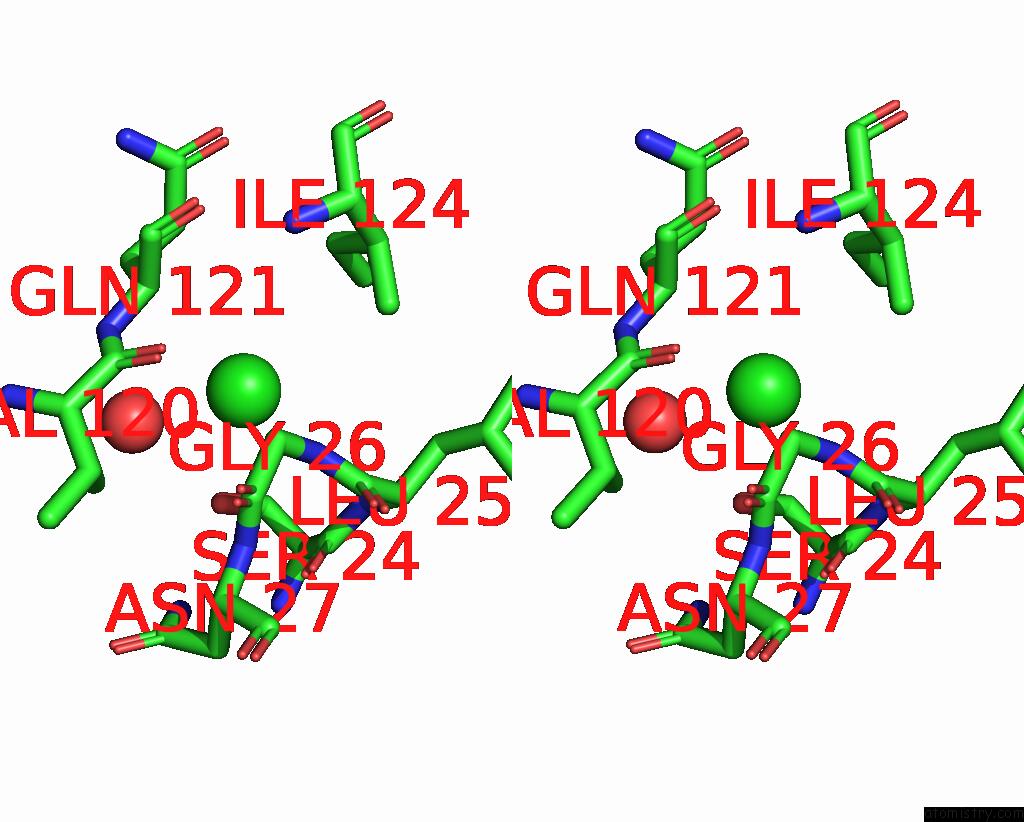
Chlorine binding site 3 out of 5 in 7ynv
Go back to
Chlorine binding site 3 out
of 5 in the Crystal Structure of Photolysed Hen Egg White Lysozyme Introduced with O-(2-Nitrobenzyl)-L-Tyrosine
Mono view
Stereo pair view
Mono view
Stereo pair view
A full contact list of Chlorine with other atoms in the Cl binding
site number 3 of Crystal Structure of Photolysed Hen Egg White Lysozyme Introduced with O-(2-Nitrobenzyl)-L-Tyrosine within 5.0Å range:
|
Chlorine binding site 4 out of 5 in 7ynv
Go back to
Chlorine binding site 4 out
of 5 in the Crystal Structure of Photolysed Hen Egg White Lysozyme Introduced with O-(2-Nitrobenzyl)-L-Tyrosine
Mono view

Stereo pair view
Mono view
Stereo pair view
A full contact list of Chlorine with other atoms in the Cl binding
site number 4 of Crystal Structure of Photolysed Hen Egg White Lysozyme Introduced with O-(2-Nitrobenzyl)-L-Tyrosine within 5.0Å range:
|
Chlorine binding site 5 out of 5 in 7ynv
Go back to
Chlorine binding site 5 out
of 5 in the Crystal Structure of Photolysed Hen Egg White Lysozyme Introduced with O-(2-Nitrobenzyl)-L-Tyrosine
Mono view

Stereo pair view
Mono view
Stereo pair view
A full contact list of Chlorine with other atoms in the Cl binding
site number 5 of Crystal Structure of Photolysed Hen Egg White Lysozyme Introduced with O-(2-Nitrobenzyl)-L-Tyrosine within 5.0Å range:
|
Reference:
T.Hosaka,
K.Katsura,
Y.Ishizuka-Katsura,
K.Hanada,
K.Ito,
Y.Tomabechi,
M.Inoue,
R.Akasaka,
C.Takemoto,
M.Shirouzu.
Crystal Structure of An Archaeal Tyrosyl-Trna Synthetase Bound to Photocaged L-Tyrosine and Its Potential Application to Time-Resolved X-Ray Crystallography. Int J Mol Sci V. 23 2022.
ISSN: ESSN 1422-0067
PubMed: 36142308
DOI: 10.3390/IJMS231810399
Page generated: Tue Jul 30 06:01:29 2024
ISSN: ESSN 1422-0067
PubMed: 36142308
DOI: 10.3390/IJMS231810399
Last articles
Zn in 9JYWZn in 9IR4
Zn in 9IR3
Zn in 9GMX
Zn in 9GMW
Zn in 9JEJ
Zn in 9ERF
Zn in 9ERE
Zn in 9EGV
Zn in 9EGW